- Visibility 78 Views
- Downloads 12 Downloads
- DOI 10.18231/j.jdp.2023.018
-
CrossMark

Rare bilateral incisiform supernumerary teeth: A case report
- Author Details:
-
 Madhusudhan K S *
-
Priya Subramaniam
-
Anushree S Shenoy
Introduction
Odontogenesis is a complex biological process involving systematic cascading interactions between epithelial component and mesenchymal component, which are the branchial arch ectoderm and the ectomesenchyme respectively.[1] Tooth number, shape and size of teeth are determined by the initiation and morphogenetic stages of tooth development.
Molecular evidence of replication signals throughout initiation and morphogensis is reflected clinically in the associations with abnormalities in number, size and shape.[2] Discrepancies in the number of teeth that develop are frequent. The present case is a 7-year-old boy with non-syndromic bilateral supplemental tooth in primary dentition.
Case Presentation
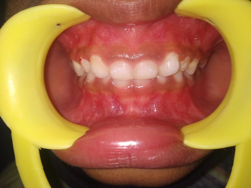
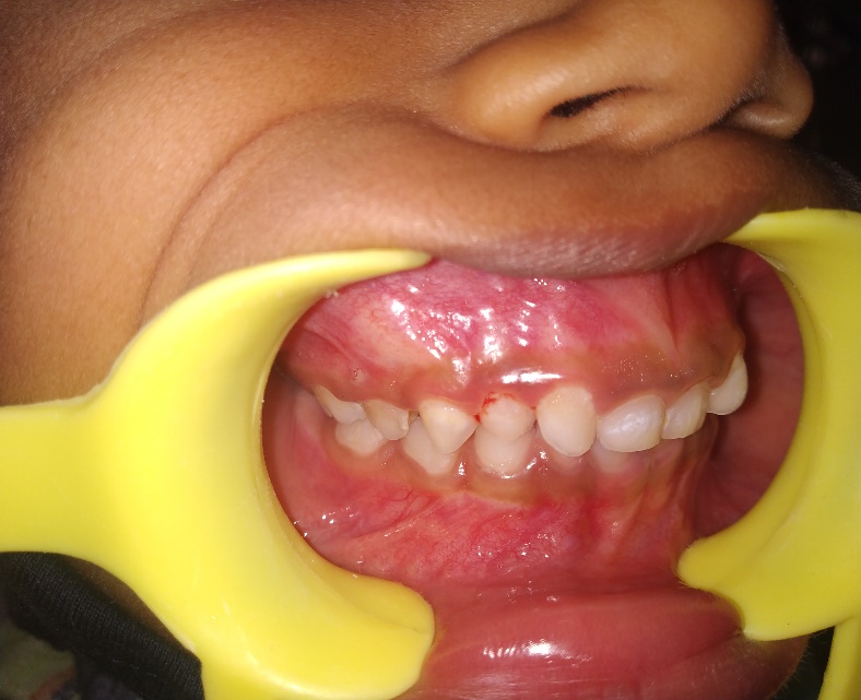
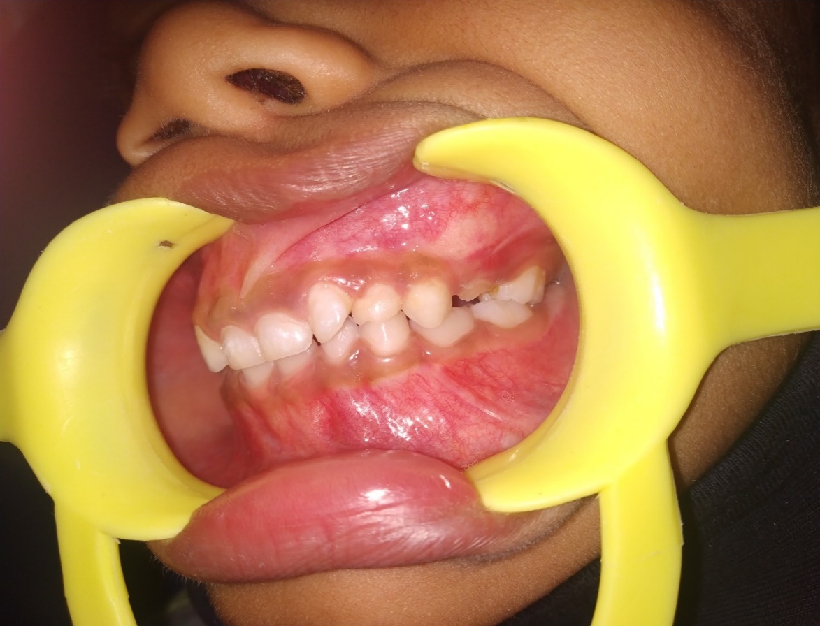
A 7-year-old boy visited the department of Pediatric and Preventive Dentistry for routine dental check-up. Medical, family, and dental history were non-contributory. General and extra oral examinations were negative. Intraoral examination revealed presence of an extra tooth which mimicked primary lateral incisors in their appearance on either side of maxillary quadrants ([Figure 1], [Figure 2], [Figure 3], [Figure 4]). Bilateral supplemental teeth are identified according to their morphology. Dental caries in relation to 55,54, 65 and gross decay in relation to 64 was noted ([Figure 4]). Based on the intra oral periapical - radiographs, bilateral supernumerary teeth mesial to primary canine was diagnosed ([Figure 6], [Figure 7]). Based on clinical findings and radiographic interpretations, the final diagnosis is bilateral supplemental teeth mesial to the maxillary primary canines. Since dental abnormalities had not compromised his oral health, preventive and restorative procedures were completed and regular follow ups are planned to intervene at appropriate intervention time.


Discussion
Supernumerary teeth are the extra teeth in normal set of dentition. They closely resemble to the group of teeth to which it belongs or may bear little resemblance in size or shape to the teeth with which it is associated. Supernumerary teeth are classified as mesiodens, para-molars, distomolar and para-premolars according to the location. Based on the morphology, it is classified as conical, tuberculate, supplemental, odontome. [3] This report describes the supplemental type of supernumerary teeth.
The incidence of supernumerary teeth is 0.8% for primary dentition and 2.1 % for permanent dentition. A possible explanation for the low incidence could be less detection by parents, reasonable alignment of supernumerary tooth or teeth due to spaced dentition. Most children have their first dental check-up after the permanent anterior teeth erupts and hence, primary anterior supernumerary teeth which have erupted and exfoliate normally often go unnoticed. [4]
A cross-sectional study of 4180 South Indian children reported a prevalence rate of 0.23% with male predominance (3:2), with common location being the right maxillary arch associated with central and lateral incisors. [5] They may occur as single or multiple, unilateral or bilateral, erupted or impacted and in one or both jaws. The most common supernumerary tooth is the mesiodens.
Multiple supernumerary teeth are often associated with syndromes. Commonly associated syndromes are: Gardiner's syndrome, Cleidocranial dysplasia, cleft lip and palate, [3] Apert syndrome, Crouzon disease, Down's syndrome, [6], [7] Chondroectodermal dysplasia, [8] familial adenomatous polyposis, trichorhinophalangeal syndrome type-1, Rubinstein-Taybi syndrome, Nance-Horan syndrome, Opitz G/BBB syndrome, oculofaciocardiodental syndrome, Robinow syndrome. Kreiborg- Pakistani syndrome, Ellisvan Creveld syndrome, Goldenhar syndrome, Noonan syndrome. [9]
In similar cases, supplemental teeth have been associated with fusion [10] and have been reported to be present in both dentitions. [6] The exact etiology is unknown and does not support the Mendelian pattern. [3] The most accepted etiology is hyperactive dental lamina, other suggested etiologices are: atavism, dichotomy of tooth germ, [11], [1] genetic and multifactorial. The dental lamina hyperactivity theory states that a supplemental form develops from the lingual extension of an accessory tooth bud whereas a rudimentary form would develop from the proliferation of the epithelial remnants of the dental lamina. [12]
Supernumerary teeth have various effects on developing dentition causing malocclusion. It can cause crowding, diastema, delayed eruption or retention, ectopic eruption, delayed or abnormal root formation, resorption of adjacent teeth, deformity and/or loss of vitality in adjacent teeth, and cysts. [13] Therefore, early intervention is as important as the prevention for permanent dentition.
The treatment options for a supernumerary tooth includes: [14]
In asymptomatic cases it is desirable to wait and watch.
Extraction followed by wait and watch.
Extraction followed by orthodontic treatment.
Conclusion
A rare case of bilateral supplemental teeth mesial to maxillary primary canines is reported. The care takers of the child were informed about the additional tooth, and the need for subsequent treatment of the carious teeth. Since orthodontic treatment has not been determined precisely and has no associated pathology, there is no need for the supplemental tooth extraction.
Declaration of Patient Consent
Parent's written consent for participation and consent is available upon request.
Source of Funding
None.
Conflict of Interest
None.
References
- GS Kumar. Orban’s Oral Histology and Embryology, 15th Edn.. 2019. [Google Scholar]
- AH Brook, J Jernvall, RN Smith, TE Hughes, GC Townsend. The dentition: the outcomes of morphogenesis leading to variations of tooth number, size and shape. Aust Dent J. 2014 2014. [Google Scholar] [Crossref]
- WG Shafer, MK Hine, BM Levy. . A Textbook of Oral Pathology. 4th Edn. 1963. [Google Scholar]
- RN Bahadure, N Thosar, Kharabe V Jain, R Gaikwad. Supernumerary Teeth in Primary Dentition and Early Intervention: A Series of Case Reports. Case Rep Dent 2012. [Google Scholar] [Crossref]
- G Shilpa, N Gokhale, S K Mallineni, S Nuvvula. Prevalence of dental anomalies in deciduous dentition and its association with succedaneous dentition: A cross-sectional study of 4180 South Indian children. J Indian Soc Pedod Prev Dent 2017. [Google Scholar]
- J F Zhu, M Marcushamer, DL King, RJ Henry. Supernumerary and congenitally absent teeth: a literature review. J Clin Pediatr Dent 1996. [Google Scholar]
- BL Jensen, S Kreiborg. Development of the dentition in cleidocranial dysplasia. J Oral Pathol Med 1990. [Google Scholar]
- G Yildirim, S Bayrak. Early diagnosis of bilateral supplemental primary and permanent maxillary lateral incisors: a case report. Eur J Dent 2011. [Google Scholar]
- F Cammarata-Scalisi, A Avendaño, M Callea. Main genetic entities associated with supernumerary teeth. Arch Argent Pediatr 2018. [Google Scholar]
- F Ghaderi, A Rafice. Bilateral Supernumerary Deciduous Maxillary Lateral Incisors with Fusion: Report of a Rare Case. J Dent (Shiraz) 2016. [Google Scholar]
- LD Rajab, MA Hamdan. Supernumerary teeth: review of the literature and a survey of 152 cases. Int J Paediatr Dent 2002. [Google Scholar]
- NM King, S Tongkoom, A Itthagarun, HM Wong, CK Lee. A catalogue of anomalies and traits of the primary dentition of southern Chinese. J Clin Pediatr Dent 2008. [Google Scholar]
- A Högström, L Andersson. Complications related to surgical removal of anterior supernumerary teeth in children. ASDC J Dent Child 1987. [Google Scholar]
- MT Garvey, HJ Barry, M Blake. Supernumerary teeth--an overview of classification, diagnosis and management. J Can Dent Assoc 1999. [Google Scholar]