- Visibility 166 Views
- Downloads 8 Downloads
- DOI 10.18231/j.jdp.2023.024
-
CrossMark

Perks of intraoral scanner to prosthodontist: A review
Introduction
The use of CAD/CAM in dentistry was first introduced by Dr. Francois Duret in 1973.[1] Intraoral scanners (IOS) have been boon to dentistry since 1980 as it has variety of applications such as impression making, use in prosthodontics and orthodontics appliances, retainers.[2], [3] The use of Intraoral Scanners (IOS) has been increased since last few years replacing the conventional impression techniques. Intraoral scanning is advanced technology used along with additive manufacturing such as 3-D printing and subtractive technology such as CAD/CAM. Nowadays Intraoral Scanners (IOS) and CAD/CAM are very popular in almost every branch of dentistry like prosthodontics, orthodontics, conservative dentistry, maxillofacial surgery.[4]
The drawbacks of conventional impression techniques such as volumetric changes of impression materials and expansion of dental stone have been compensated by Intraoral Scanners (IOS). [5] Intraoral Scanners (IOS) and CAD/CAM provide better planning of treatment, case acceptance, communication with laboratories, less clinical time, storage requirements, and less treatment times. [6] So, the aim of current study was to:
To know about intraoral scanners
Benefits and limitations of Intraoral Scanners (IOS)
Workflow of Intraoral Scanners (IOS).
Various Commercial Intraoral Scanners (IOS) available in market.
Principle of Intraoral Scanners (IOS)
Digital intraoral scanner is divided into following parts: image capture, data processing, and onscreen results. The primary factor is the first factor i.e., image capture. The principle on which Intraoral Scanners (IOS) works are as follows:
Confocal laser scanning: The LASER is directed toward the object through a narrow hole. The area which is out of focus is not recorded. The 3D image is created by merging different 2D confocal planes. Thus, it is also called as “point and stitch technique”
Triangulation technique: It consists of three points: - the LASER, sensor and object surface. The information of the object is gained through Pythagorean theorem.
Active wave-front sampling (3D-in-motion video recording): In this the 3D information of object surface is collected by single lens imaging system. A radio-opaque powder can be sprinkled before scanning for greater accuracy. [1]
Stereophotogrammetric technology: First of all, a stereo camera with an infrared flash detects the position of special flag abutments screwed into the implants. It is based upon registering the x, y and z coordinates of each implant and the distances between them. This information is then converted into a stereolithographic (STL) file. [7], [8], [9], [10] To add the soft-tissue information, the user must obtain another STL file by using an intraoral or extraoral scanner. [11] Stereophotogrammetry estimates all coordinates (x, y, and z) only through an algorithmic analysis of image. As this approach relies on passive light projection and software rather than active projection and hardware, the camera is relatively small, its handling is easier, and its production is cheaper. [6]
Reconstruction Technologies: The most difficult challenge of generating a 3D numerical model is the matching of POI taken under different angles. [2], [3], [12] Distances between different pictures can be calculated using an accelerometer incorporated in the camera, but a similarity calculation is more often used to determine the point of view of the image. [6]
Benefits of intraoral scanners (IOS)
Patient Comfort: Conventional impression techniques may cause discomfort to the patient such as severe gag reflex with impression material, position of impression tray. These all problems are compensated in Intraoral Scanners (IOS). Intra oral scanners have eliminated the need of conventional impression materials and trays which are often disliked by patient. It has seen in many surveys that patient prioritize the digital impressions over conventional impressions. [13]
Reduced Clinical and laboratory time: The chairside time has been reduced with the use of digital intra oral scanners as compared to conventional impression procedures. In digital impressions the need for pouring cast after impression is eliminated. As well as the there is no need to send the cast to the lab via courier as digital cast can be sent via STL file. [13]
Easier communication with patients: Digital impressions makes the communication easier with the patient. Also, it helps in marketing. Patient feels more involved in this treatment, ultimately having powerful impact on treatment. [13]
Disadvantages of intraoral scanners (IOS)
Learning curve: It is easier for new generation dentists to get adopted to Digital Intraoral Scanners (IOS). While Older dentists find it difficult to use Intraoral Scanners (IOS). In market there are variety of scanners available which would produce different results. With advent of technology there are new Intraoral Scanners (IOS) coming in market with newer technology. [13]
Expensive assembly and cost of treatment: The cost of digital Intraoral Scanners (IOS) is very much higher than conventional impression techniques. Also, the maintenance cost of digital Intraoral Scanners (IOS) is higher. So, patient cannot afford such higher cost for treatment. [13]
Inability to detect deeper areas to prepared teeth: Most commonly occurred problem with digital Intraoral Scanners (IOS) is inability to detect deeper areas such as finish line on prepared teeth. It occurs mainly in cases of subgingival margins. Subgingival margins are given mainly in anterior region where aesthetic is more concerned but Intraoral Scanners (IOS) will find it difficult to detect subgingival margins as light may not reach that area correctly. [13]
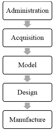
Workflow
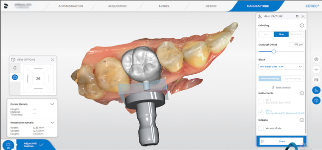
The workflow followed by omnicam scanner involves following phases ([Figure 1], [Figure 4])
Administration: In this phase all the patient’s detail are entered such as Patient’s name, Date of birth, Clinician’s name.
Acquisition: In this phase scanning of the prepared tooth and arch is done. Scanning of opposing arch as well as buccal scan is also done.
Model: Model is prepared, trimmed and edited from scanning.
Design: Design for the desired preparation is done according to Clinicians choice.
Manufacturing: From above all data the prosthesis is manufactured by milling and sintering.
Various Commercial Intraoral Scanners (IOS) Available in Market
iTero element - Align tech
It works on the principle confocal laser scanning. There is no need of powder for scanning. And it scans the arch by directly touching the tooth. The dimensions of Wand tip are 69.8 X 53.5 X 338.5 mm which is comparatively larger than other scanners. This intra oral scanner has reflective mirror into its wand thus it becomes easier for distal tooth scanning. [14], [15] The files of this scanner can be exported as STL file. The capture speed of this scanner is 6000 images/sec. [1]
Lava COS – 3M
The imaging principle on which this scanner works is active wave-front sampling technology. In this the 3D information of object surface is collected by single lens imaging system. A radio-opaque powder can be sprinkled before scanning for greater accuracy [1]. This scanner came into the market in year 2008. [1]
Trios3 - 3Shape
This scanner was released in 2000. This manufacturer has intraoral as well as extraoral scanner. It works on the principle of optimal sectioning and confocal laser scanning. The capture speed of this scanner is 3000 images/sec. For scanning it directly touches the tooth. The dimensions of Wand tip are 20 X 21 X 276 mm. the wand weight is 340g and the screen size is 19’’. The files of this scanner can be exported as STL file or Dicom file. [1]
True definition – 3M
The manufacturer of this scanner is 3M and it is the next version of Lava COS. The accuracy of this scanner is higher but sprinkling of powder is necessary. The imaging principle on which this scanner works is active wave-front sampling technology like its predecessor Lava COS. The dimensions of Wand tip are 14.2 X 16.2 X 254 mm. For scanning there is no need of direct contact with the tooth. It can be kept at the distance of 0-17 mm. It does not offer the color scan. [1]
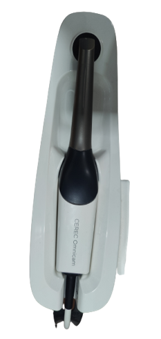
CEREC omnicam – Dentsply sirona
The imaging principle on which it works is Triangulation technique. It consists of three points the LASER, sensor and object surface. It is based upon continuous video streaming rather than stitching of static images. For scanning there is no need of direct contact with the tooth. It can be kept at the distance of 0-15 mm. The dimensions of Wand tip are 121 X 35 X 47. The wand weight is 313g. [1]
CEREC primescan – Dentsply sirona
The scan procedure is based on dynamic depth scan up to 20mm. For scanning there is no need of direct contact with the tooth. There is no need of powder for scanning. The overall dimensions are 50.9 x 58.8 x 253 mm. Full arch scanning takes 2-3 minutes. It has three sleeves: Autoclavable steel sleeve with single-use window, Single-use sleeve, Steel sleeve with sapphire glass window. [16], [17], [18], [19]
|
|
Primescan |
Omnicam |
|
Noise |
Less noisy |
Makes more noise |
|
Accuracy |
Provides more local details |
Less accurate than primescan |
|
Trueness |
Better |
Lesser than primescan but more than clinically acceptable limit. |
|
Technique sensitivity |
Less |
More |
|
Scanner head size |
15 X 15 (mm) |
10 X 10 (mm) |
|
Speed |
Faster |
Slower than primescan |
Conclusion
Digital impressions have many benefits over conventional impressions. The chief advantage is that reduced patient discomfort. Many patient factors such as anxiety and gag reflex are reduced with the help of intra oral scanners. [20], [21], [2] Intraoral Scanners (IOS) improve the patient communication. There are variety of Intraoral Scanners (IOS) in market available with several advantages over each other. [13], [22], [23], [24], [25] A complete knowledge of the Intraoral Scanners (IOS) is required for clinician to have a successful clinical strategy for scanning of prepared teeth. [6]
Source of Funding
None.
Conflict of Interest
There is no conflict of interest
References
- H Hann-Min Hwang, CW Chou, YJ Chen, CCJ Yao. An Overview of Digital Intraoral Scanners: Past, Present and Future- From an Orthodontic Perspective. Taiwanese J Orthod 2018. [Google Scholar] [Crossref]
- T Joda, P Lenherr, P Dedem, I Kovaltschuk, U Bragger, NU Zitzmann. Time efficiency, difficulty, and operator's preference comparing digital and conventional implant impressions: a randomized controlled trial. Clin Oral Implants Res 2017. [Google Scholar] [Crossref]
- S Amin, H P Weber, M Finkelman, K El Rafie, Y Kudara, P Papaspyridakos. Digital vs. conventional full-arch implant impressions: a comparative study. Clin Oral Implants Res 2017. [Google Scholar] [Crossref]
- V Daniel, D Banyai, G Zoltan, J Borbely, P Hermann, T Hegedüs. User Experience of Intraoral Scanners in Dentistry: Transnational Questionnaire Study. Int Dent J 2023. [Google Scholar] [Crossref]
- B Giménez, M Özcan, F Martínez-Rus, G Pradíes. Accuracy of a digital impression system based on active wavefront sampling technology for implants considering operator experience, implant angulation, and depth. Clin Implant Dent Relat Res 2015. [Google Scholar] [Crossref]
- R Richert, A Goujat, L Venet, G Viguie, S Viennot, P Robinson. Intraoral Scanner Technologies: A Review to Make a Successful Impression. J Healthc Eng 2017. [Google Scholar] [Crossref]
- KM Chochlidakis, P Papaspyridakos, A Geminiani, CJ Chen, IJ Feng, C Ercoli. Digital versus conventional impressions for fixed prosthodontics: A systematic review and meta-analysis. J Prosthet Dent 2016. [Google Scholar]
- C R Means, I E Flenniken. Gagging-a problem in prosthetic dentistry. J Prosthet Dent 1970. [Google Scholar]
- P Rosted, M Bundgaard, J Fiske, A M Pedersen. The use of acupuncture in controlling the gag reflex in patients requiring an upper alginate impression: an audit. Br Dent J 2006. [Google Scholar]
- JD Muir, EJ Calvert. Vomiting during the taking of dental impressions. Two case reports of the use of psychological techniques. Br Dent J 1988. [Google Scholar]
- G Pradíes, A Ferreiroa, M Özcan, B Giménez, F Martínez-Rus. Using stereophotogrammetric technology for obtaining intraoral digital impressions of implants. J Am Dent Assoc 2014. [Google Scholar] [Crossref]
- A Marghalani, H P Weber, M Finkelman, Y Kudara, K El Rafie, P Papaspyridakos. Digital versus conventional implant impressions for partially edentulous arches: An evaluation of accuracy. J Prosthet Dent 2018. [Google Scholar] [Crossref]
- F Mangano, A Gandolfi, G Luongo, S Logozzo. Intraoral scanners in dentistry: a review of the current literature. BMC Oral Health 2017. [Google Scholar] [Crossref]
- GJ Christensen. Will digital impressions eliminate the current problems with conventional impressions?. J Am Dent Assoc 2008. [Google Scholar] [Crossref]
- E Yuzbasioglu, H Kurt, R Turunc, H Bilir. Comparison of digital and conventional impression techniques: evaluation of patients' perception, treatment comfort, effectiveness and clinical outcomes. BMC Oral Health 2014. [Google Scholar] [Crossref]
- M Imburgia, S Logozzo, U Hauschild, G Veronesi, C Mangano, F G Mangano. Accuracy of four intraoral scanners in oral implantology: a comparative in vitro study. BMC Oral Health 2017. [Google Scholar] [Crossref]
- ML Aragón, LF Pontes, LM Bichara, C Flores-Mir, D Normando. Validity and reliability of intraoral scanners compared to conventional gypsum models measurements: a systematic review. Eur J Orthod 2016. [Google Scholar]
- C Goracci, L Franchi, A Vichi, M Ferrari. Accuracy, reliability, and efficiency of intraoral scanners for full-arch impressions: a systematic review of the clinical evidence. Eur J Orthod 2016. [Google Scholar]
- P Ahlholm, K Sipilä, P Vallittu, M Jakonen, U Kotiranta. Digital Versus Conventional Impressions in Fixed Prosthodontics: A Review. J Prosthodont 2016. [Google Scholar] [Crossref]
- R Nedelcu, P Olsson, I Nystrom, J Ryden, A Thor. Accuracy and precision of 3 intraoral scanners and accuracy of conventional impressions: A novel in vivo analysis method. J Dent 2018. [Google Scholar] [Crossref]
- T Joda, U Bragger. Time-efficiency analysis comparing digital and conventional workflows for implant crowns: a prospective clinical crossover trial. Int J Oral Maxillofac Implants 2015. [Google Scholar]
- C B Martin, E V Chalmers, G T Mcintyre, H Cochrane, P A Mossey. Orthodontic scanners: what's available?. J Orthod 2015. [Google Scholar]
- T Joda, U Brägger. Patient-centered outcomes comparing digital and conventional implant impression procedures: a randomized crossover trial. Clin Oral Implants Res 2016. [Google Scholar]
- M A Atieh, A V Ritter, C C Ko, I Duqum. Accuracy evaluation of intraoral optical impressions: A clinical study using a reference appliance. J Prosthet Dent 2017. [Google Scholar] [Crossref]
- C Goracci, L Franchi, A Vichi, M Ferrari. Accuracy, reliability, and efficiency of intraoral scanners for fullarch impressions: a systematic review of the clinical evidence. Eur J Orthod 2016. [Google Scholar] [Crossref]
- Introduction
- Principle of Intraoral Scanners (IOS)
- Workflow
- Various Commercial Intraoral Scanners (IOS) Available in Market
- iTero element - Align tech
- Lava COS – 3M
- Trios3 - 3Shape
- True definition – 3M
- CEREC omnicam – Dentsply sirona
- CEREC primescan – Dentsply sirona
- Conclusion
- Source of Funding
- Conflict of Interest