- Visibility 669 Views
- Downloads 47 Downloads
- Permissions
- DOI 10.18231/j.jdp.2021.025
-
CrossMark

Clinical evaluation of excisional new attachment procedure versus laser assisted new attachment procedure in the treatment of periodontal pockets
Abstract
Introduction: The Laser Assisted New Attachment Procedure (LANAP) protocol is a laser based periodontal regenerative procedure and was patterned, conceptually, after the Excisional New Attachment Procedure (ENAP) to separate the diseased epithelium from the underlying connective tissue and to selectively vaporize and disrupt inflamed and necrotic tissue from connective tissue.
Objective: The present study was designed to compare and evaluate the clinical efficacy of LANAP versus ENAP in the treatment of chronic periodontitis.
Materials and Methods: Twenty periodontal pocket sites of ≥5mm in systemically healthy patients were selected and randomly allocated to either (ENAP) Excisional New Attachment Procedure (Group I) or (LANAP) Laser Assisted New Attachment Procedure (Group II). Patients were evaluated for Plaque Index, Gingival Index, Gingival Bleeding Index, Probing Depth, Loss of attachment, recession and VAS scores. Patients were recalled for follow up at 1 week, 1 month and 3 months at which clinical parameters were recorded.
Results: The results of the present study revealed statistically insignificant difference in both groups however clinically significant reduction in probing depth was seen with ENAP as compared to LANAP but laser procedure had less VAS scores as compared to ENAP.
Conclusion: Although probing depth reduction was more for ENAP, patient discomfort with less bleeding was observed with LANAP.
Introduction
Periodontitis is a multifactorial inflammatory disease of periodontal tissue caused by extension of bacterial infection into the gingiva,[1] which leads to connective tissue destruction and alveolar bone loss.[2], [3] The essential objective of periodontal treatment is to decrease or eliminate the responsible periodontal pathogens,[4] by means of removing bacterial deposits from the tooth surface.[5]
Conventional mechanical debridement (scaling and root planning) is considered to be the gold standard for inflammatory periodontal disease. [6] The ultimate goal of periodontal therapy is the formation of a new connective tissue attachment & re-growth of alveolar bone. This new connective tissue attachment can only be achieved when the epithelial migration can be prevented on the treated root surface. Over the years, various modes of therapy have been suggested to avoid epithelial migration, which includes soft tissue curettage, various types of flap procedures including Modified Widman flap, Guided Tissue Regeneration and, more recently Lasers.[7]
The Excisional New Attachment Procedure has been described as “curettage with a scalpel”. The objective of the excisional new attachment procedure is to provide easy access to the root surface for visual treatment. The epithelium of the soft pocket wall is excised by means of a reverse bevel incision, and a mucosal flap is reflected without exposing the alveolar bone, to allow for access to the root surface. The mucosal flap is then readapted in its original position with interdental sutures. The primary goal of this surgery is the removal of pocket epithelium.[8]
Lasers are one of the most promising new technical modalities for non- surgical periodontal treatment for achieving excellent tissue ablation with strong bactericidal and detoxification effects. [9] LANAP is “cementum-mediated new attachment to the root surface in the absence of a long junctional epithelium.” LANAP utilizes a free-running (10- 6 seconds) pulsed Nd:YAG laser in place of a scalpel. A thin 0.3 to 0.4 laser fiber permits easy access deep into the periodontal pocket without the need to surgically elevate a flap.[10] Laser treatment is expected to promote healing of periodontal tissue, ablating the inflamed lesion and epithelial lining of soft tissue wall within periodontal pockets. This procedure might be more effective for the treatment of residual pockets after initial therapy and during maintenance. Part of the laser energy scatters and energy level might then stimulate the cells of surrounding tissue, resulting in reduction of inflammatory conditions, in cell proliferation, and increased flow of lymph improving the periodontal tissue attachment and possibly reducing postoperative pain.[9]
Aim
The present study was designed to compare and evaluate the clinical efficacy of diode laser as an adjunct to SRP versus ENAP in the treatment of chronic periodontitis.
Materials and Methods
Twenty periodontal pocket sites of ≥5mm in systemically healthy patients were selected from the Out Patient Department of Periodontology, Institute of Dental Studies & Technologies, Modinagar, UP. All the patients underwent scaling and root planing and thereafter were randomly allocated to either Excisional New Attachment Procedure (Group I) or Laser Assisted New Attachment Procedure (Group II).
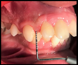
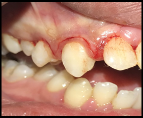
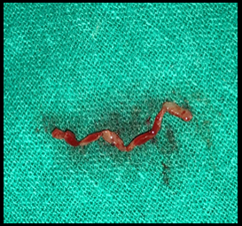
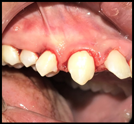
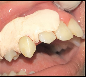
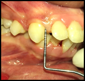
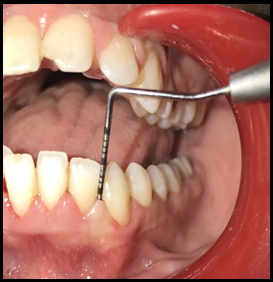
In Group I Excisional New Attachment Procedure (10 sites), after infiltrating the area with local anesthesia, depth of the pocket was marked with the help of a pocket marker ([Figure 1]) then with No.15 or 11 scalpel blade scalloped reverse bevel incision was made from the crest of gingiva to the base of sulcus ([Figure 2]). The inflamed granulated and excised tissues was removed with the help of scalers and curettes ([Figure 3]). The root was scaled hard and smooth and was free of calculus and the area was flushed with normal saline to remove debris, blood clots and tissue tags ([Figure 4]). Sutures and periodontal pack were placed if necessary ([Figure 5]).
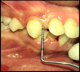
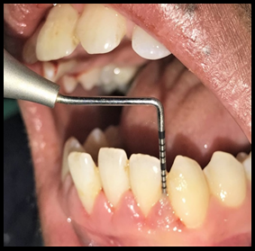
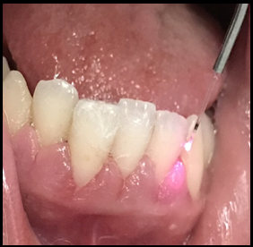
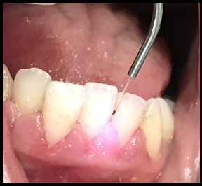
In Group II Laser Assisted New Attachment Procedure (10 sites), bone sounding under anesthesia was done to identify bone defects depth ([Figure 6]). 810 nm wavelength and 1.0 W/cm2 power density diode laser tip was inserted into the pockets (3 cycles each of 10 seconds duration and interrupted for 10 seconds each) ([Figure 7]). The tip was constantly moved with the objective to remove epithelial lining ([Figure 8]). Patients were recalled for follow up at 1 week, 1 month and 3 months at which clinical parameters were recorded.
Clinical parameters (Plaque Index, Gingival Index, Gingival Bleeding Index, Probing Depth, Loss of Attachment, Recession) were recorded. Plaque index, Gingival Index, Gingival Bleeding Index were recorded on the day of surgery (Baseline), 1 week and 1 month. Probing Depth, Loss of Attachment, Recession were recorded on the day of surgery (Baseline) and 1 month.
Descriptive statistics was performed by calculating mean and standard deviation for the continuous variables. Categorical variables are presented as absolute numbers and percentage. Nominal categorical data between the groups were compared using chi-square goodness-to-fit test. The collected data was tabulated and subjected to statistical analysis. The software used for the statistical analysis were SPSS (statistical package for social sciences) version 25.0 and MedCalc software. The following results were obtained.
Group I- Excisional new attachment procedure
Group II- Laser assisted new attachment procedure([Figure 7], [Figure 8], [Figure 9], [Figure 10], [Figure 11])
Results
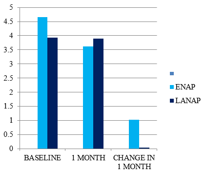
The result of the present study revealed that during intragroup comparison there was statistically significant reduction in mean values of PI, GI, BOP, PPD and CAL in both ENAP and LANAP groups when compared from baseline to follow up visits ([Figure 11], [Figure 12], [Figure 13], [Figure 14], [Figure 15]).
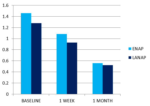
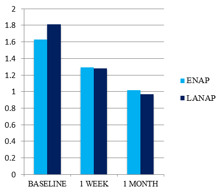
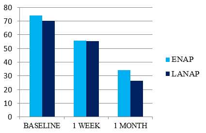
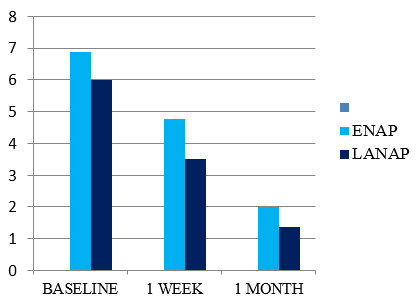
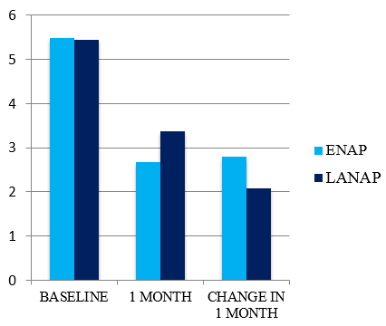
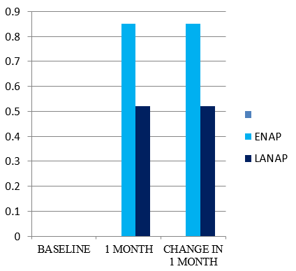
During intergroup comparisons PI and GI showed statistically insignificant differences in both the groups ([Table 1] ). Clinically significant reduction in probing depth was seen more with ENAP as compared to LANAP. Also GR and CAL showed clinically insignificant differences in both the groups (Figure16 and 17). However laser procedure had less BOP and VAS score as compared to ENAP.
|
|
ENAP v/s LANAP (p-value) |
|
|
|
Change in 1 week |
Change in 1 month |
|
PI |
0.560 |
0.173 |
|
GI |
0.049 |
0.029 |
|
BOP |
0.562 |
0.560 |
|
PPD |
- |
0.001 |
|
GR |
- |
0.088 |
|
CAL |
- |
0.017 |
|
VAS |
0.462 |
0.666 |
Discussion
Present study revealed that during intragroup comparison there was statistically significant reduction in mean values of PI, GI, BOP, PPD and CAL in both ENAP and LANAP groups when compared from baseline to follow up visits. During intergroup comparisons PI and GI showed statistically insignificant differences in both the groups. Clinically significant reduction in probing depth was seen more with ENAP as compared to LANAP. The greater reduction of PD in ENAP group may be due to complete removal of the sulcular epithelium and the possibility of gaining soft tissue attachment to the tooth. Histological studies have confirmed that this attachment is most likely to be a long junctional epithelium. These findings was in accordance with the studies done by Raymond Yukna, Ramfjord et al and Zamet et al. Lobo and Paul in their study concluded that the use of diode laser for open flap debridement did not significantly benefit the treatment outcome. Also GR and CAL showed clinically insignificant differences in both the groups. However laser procedure had less BOP and VAS score as compared to ENAP these findings was in accordance with study done by Harshada et al. Conlan found laser therapy of periodontal pockets does not involve discomfort or intraoperative pain, nor requires, the execution of locoregional anesthesia since the power values provided are relatively low and the energy is supplied in pulsed mode. Also study done by Katuri indicates that the LANAP showed decreased gingival bleeding and less pain as laser therapy blocks the pain signal transmitted from the injured parts of the body to the brain. This decrease nerve sensitivity and significantly reduces the perception of pain. With the diode laser there is reduced need for systemic or locally applied antimicrobials.
Conclusion
Although probing depth reduction was more for ENAP, patient discomfort with less bleeding was observed with LANAP.
Conflict of Interest
The authors declare that there are no conflicts of interest in this paper.
Source of Funding
None.
References
- Andersen R, Loebel N, Hammond D, Wilson M. Treatment of periodontal disease by photodisinfection compared to scaling and root planing. J Clin Dent. 2007;18(2):34-8. [Google Scholar]
- Renvert S, Dahlen G, Wikstrom M. Treatment of periodontal disease based on microbiological diagnosis. Relation between microbiological and clinical parameters during 5 years. J Periodontol. 1996;67(6):562-71. [Google Scholar]
- Sbordone L, Ramaglia L, Gulletta E, Iacono V. Recolonization of the subgingival microflora after scaling and root planing in human periodontitis. J Periodontol. 1990;61(9):579-84. [Google Scholar] [Crossref]
- Teles RP, Haffajee AD, Socransky SS. Microbiological goals of periodontal therapy. Periodontol. 2000;42:180-218. [Google Scholar] [Crossref]
- Cobb CM. Non-surgical pocket therapy: mechanical. Ann Periodontol. 1996;1(1):443-90. [Google Scholar] [Crossref]
- Soukos NS, Goodson JM. Photodynamic therapy in the control of oral biofilms. Periodontol. 2000;55(1):143-66. [Google Scholar] [Crossref]
- Roopa DA, Shrinkhala S, Goswami A. LANAP- A new hope in the treatment of periodontitis. Rama Univ J Dent Sci. 2016;3:30-3. [Google Scholar]
- Adriaens PA, Adriaens LM. Effects of nonsurgical periodontal therapy on hard and soft tissues. Periodontol. 2000;36:121-45. [Google Scholar]
- Konopka K, Goslinski T. Photodynamic therapy in dentistry. J Dent Res. 2007;86(8):694-707. [Google Scholar] [Crossref]
- Harris DM, Gregg RH, Mccarthy. Laser-assisted new attachment procedure in private practice. Gen. Dent. 2004;52(5):396-403. [Google Scholar]