Introduction
The harmony of smile is determined by gingival health along with the shape, colour and position of teeth.1 Gingival health and appearance are essential components of an attractive smile. Healthy gingiva and its appearance are essential for an aesthetic smile and removal of hyperpigmentation is necessary for an attractive and beautiful smile. 2 Colour of gingiva depends on number and size of blood vessels, epithelial thickness, quantity of keratinization and pigments within epithelium like melanin, carotene, reduced haemoglobin and oxyhemoglobin. 3 Melanin is the most common natural pigment contributing to colour of gums. Although melanin pigmentation of gingiva is completely benign and does not present medical problems. Complaints of black gums are common particularly in patients having very high smile line (gummy smile). The first and foremost important indication of depigmentation is patient’s improved aesthetics.
Oral melanin pigmentation is considered to be multifactorial. It can be physiological or pathological and can be caused by a variety of local and/or systemic factors such as tobacco use, genetic, prolonged administration of certain drugs like antimalarial agents and tricyclic anti-depressants. 4
Oral pigmentation is a discoloration of the gingival/oral mucosa, associated with several exogenous and endogenous factors. Etiological factors are varied which include drugs, heavy metals, genetics, endocrine disturbances, syndromes as Albright's syndrome, Peutz Jegher's syndrome, and also in inflammation. Adverse habits such as smoking can also stimulate melanin pigmentation and the intensity of pigmentation is related to the duration of smoking and the number of cigarettes consumed. 5 It is generally agreed that pigmented areas are present only when melanin granules synthesized by melanocytes are transferred to keratinocytes. This close relation between melanocytes and keratinocytes was labelled by Fitzpatrik and Breathnach (1963) as the epidermal-melanin unit. 6 The distribution and intensity of the pigmentation varies and hence, the technique should be based on clinical experience and individual preference, which give better results. 7 Various methods such as gingivectomy with free gingival autografts and acellular dermal matrix, allograft, electrosurgery, cryosurgery, abrasion with diamond burs, scalpel and laser therapy have been used for the treatment of gingival melanin pigmentation with various degree of success. 8
Demand for cosmetic therapy of gingival melanin depigmentation is common. Gingival depigmentation has been carried out using non-surgical and surgical approaches.
Methods aimed at removing pigmented layer2
A. Surgical methods of depigmentation
B. Methods aimed at masking the pigmented gingiva with grafts from less pigmented areas.
The following is the case report of gingival depigmentation procedure performed using 980 nm diode laser. Infiltration anesthesia was given to the patient and depigmentation procedure was done.
Case Report
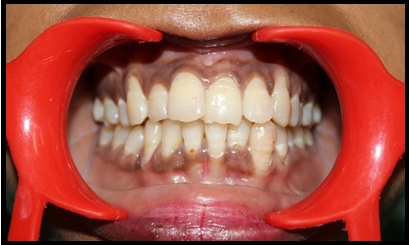
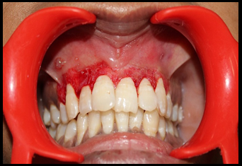
A 28 year old female reported to the outpatient department of the institute, with a chief complaint of black coloured gingiva, since she was very conscious about her esthetic appearance, she wants to get it corrected. Intra oral examination revealed deeply pigmented gingiva from canine to canine in both maxillary and mandibular arches (Figure 1). The pigmentation was esthetically displeasing and hence gingival depigmentation procedure was planned only in maxillary arch, since the mandibular region was not visible during smiling and taking etc. For right maxillary quadrant, scalpel technique for depigmentation was planned and for left maxillary quadrant, use of diode laser was planned. Accordingly, phase I therapy including thorough scaling and root planning was performed and routine blood examination of patient was done which revealed all values within the normal limits.
Procedure
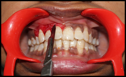
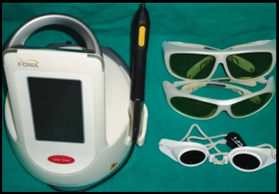
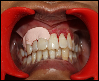
For right side, local anaesthesia was infiltrated. Bard-Parker with blade no 15 was used to remove the pigmented layer (Figure 2). Pressure was applied with a sterile gauze moistened in saline to control haemorrhage during the procedure. For left maxillary side, topical anaesthesia spray was used, Sirona laser having wavelength 810 to 980 nm at 1.5 to 2 Watt power in a continuous wave mode with a flexible fibre optic was used. (Figure 3)
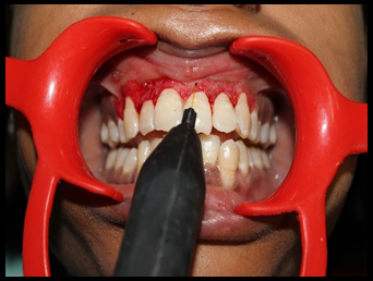
After the selected power setting were entered, the laser was activated. The procedure was performed in contact mode, with light sweeping brush strokes (Figure 4). The laser fibre optic was directed to the target tissue until white scab formation occurred. White scab gingiva was scraped off with wet saline moistened gauze to remove epithelium containing melanin pigmentation. No pain was experienced by the patient during the procedure.
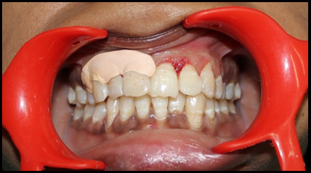
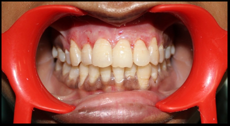
Both sides were appeared normal immediately after the surgery (Figure 5, Figure 6). Periodontal pack was given to the patient on side treated with scalpel. Patient was recalled on 3rd day. (Figure 7) Progressive healing of surgical site was seen. Patient didn’t report any pain or discomfort on side treated with laser. She felt little discomfort and pain on the side treated with scalpel. After 1 week patient was recalled for review. Wound healing was evaluated based on epithilization & Visual analog scale (Figure 8). Similarly same procedure was carried out for 10 patients.
Results
In all the above case, no postoperative pain, hemorrhage, infection or scarring occurred in any of the site where laser was used. The patients were observed at 1 day, 3 days and 1 week after the procedure and the healing was found to be uneventful. The patient’s acceptance of the procedure was good and the results were excellent, as perceived by the patient. The follow-up period spanned over 3 months. There was no recurrence of pigmentation observed in 3 months.
Discussion
Various methods of de-epithelization of the pigmented areas of the gingiva have been documented, such as scalpel surgery, gingivectomy, gingivectomy with free gingival autograft, cryosurgery, electrosurgery, chemical agents such as 90% phenol and 95% alcohol, abrasion with diamond bur, Nd:YAG laser, semiconductor diode laser and CO2.
Scalpel technique for depigmentation is the most economical one as compared to other techniques which requires more advance armamentarium. Scalpel technique is relatively simple and versatile and it requires minimum time and effort. However, scalpel surgery causes unpleasant bleeding during and after operation and it is necessary to cover the surgical site with periodontal dressing for 7 to 10 days. This technique requires good hand movement control of clinician to avoid excessive cutting of gingiva.
Electrosurgery requires more expertise than scalpel surgery. Prolonged or repeated application of current to the tissues induce heat application and undesired tissue destruction. Contact of current with the periosteum or the alveolar bone and vital teeth should be avoided. Cryosurgery procedure is followed by considerable swelling and it is also accompanied by increased soft tissue destruction. Depth control is difficult and optimal duration of freezing is not known, but prolonged freezing increases tissue destruction.
Free gingival graft is quite an invasive and an extensive procedure and it has not been advised for depigmentation procedures routinely. It also has the disadvantage of second surgical site, additional discomfort and poor tissue color matching at the recipient site.
Depigmentation was performed using 980nm diode laser. It is a one-step laser treatment and is sufficient to eliminate the pigmented areas and does not require any periodontal dressing.
Laser has the advantage of easy handling, it is less time consuming, ability to hemostasis, and effects less or no pain after the tretment, less discomfort. But laser is an expensive and sophisticated equipment that is not available commonly at all places it makes the treatment expensive.
Lasers were introduced in 1960 by Maiman and were brought into general practice by Dr William and Terry Myers. Different lasers such as carbon dioxide CO2 laser, Nd:YAG laser, semicondonductor diode laser, argon laser, Er:YAG laser and Er,Cr.YSGG laser have been reported as effective, pleasant and reliable method with minimal postoperative discomfort and faster wound healing depigmentation procedure. 4 The diode laser is a solid state semiconductor laser that typically uses a combination of gallium (Ga), Arsenide (Ar), and other elements, such as aluminium (Al) and indium (In), to change electrical energy into light energy. Dental laser energy has an affinity for different tissue components. The 980 nm diode laser has energy and wavelength characteristics that specially target the soft tissues. It has an affinity for hemoglobin and melanin, therefore it more efficient and better equipped to address deeper soft tissue problems 9 Since, the diode does not interact with dental hard tissues at reduced power settings, the laser is an excellent soft tissue surgical laser indicated for cutting and coagulating gingiva and oral mucosa, and for soft tissue curettage or sulcular debridement. There is little information on the behavior of melanocytes after surgical injury. Spontaneous repigmentation has been shown to occur and mechanism suggested is that the active melanocytes from the adjacent pigmented tissues migrate to treated areas. The large variation in time of repigmentation may be related to the technique used and the race of the patient. Repigmentation may also attribute to the melanocytes which were left during the surgery as stated by Ginwalla et al. 10 These may become activated and start synthesizing melanin.
Soft tissue lasers help in healing by reducing inflammation and pain. Its applications include: Frenectomies, Incision and Excision Biopsy, Crown lengthening, Operculum removal, Lower Level Laser Therapy LLLT for TMD pain, LLLT for Post Extraction/Impaction pain, LLLT for Post Implant pain and discomfort, Implant second stage(screw exposing), for treating Ulcers, Whitening, Endo procedures (Canal irrigation) , Perio procedures (Curettage of gums), Bleaching.
Diode laser has high electrical to optical efficiency, are small light weight and compact, hence portable and are quiet devices as compared to other solid state and gas lasers (such as Nd:YAG, KTP.YAG, HO.YAG, argon, erbium family and CO2). 11 The procedure can be performed using topical or infiltration anesthesia depending on patient’s pain threshold and the comfort of the operator.
Conclusion
The diode laser is minimally invasive treatment option for the elimination of unesthetic gingival melanin pigmentation. Scalpel procedure causes unpleasant bleeding during the procedure and requires covering the unexposed surface with periodontal dressing. Diode lasers are minimal invasise incomparison to scalpel technique and less traumatic to the patient in terms of bleeding and pain.


