- Visibility 63 Views
- Downloads 7 Downloads
- DOI 10.18231/j.jdp.2021.028
-
CrossMark
- Citation
Management of endo perio lesion with class iii furcation defect –A case report
Introduction
In 1964, Simring and Goldberg first described the relationship between periodontal and pulpal disease. [1] A significant relationship between periodontium and endodontium is due to the interlinking between tissues of periodontium (precursor of PDL) and dental pulp (precursor of dental pulp) that share a common mesodermal origin, hence, any disease or involvement of bacteria, fungi or virus on one tissue causes involvement of the other.2 Iatrogenic factors such as perforations, over-instrumentation, trauma, dental malformations, root resorption, incomplete root canal treatment, chemicals used in dentistry, over or under obturation, over-irrigation, coronal leakage, poor restorations can also cause endo-perio lesions. [2], [3] Bacteria involvement in the pulp can be transferred to the periodontium (retrograde periodontitis) through lateral canals, furcation canal or apical foramen and vice versa. [4], [5]
On visual examination, there can be swelling on the buccal aspect of the infected tooth and surrounding periodontium, abscess formation may or may not occur. The pocket depth can be measured using William’s periodontal probe, tooth mobility may or may not be present. The degree of furcation involvement on multiple rooted teeth can be measured with the help of Nabers probe. Pulp vitality tests can be done to check the response of pulp on application of stimuli. [6] The origin of lesion developed due to endodontic or periodontal involvement, degree and extent of bone loss can be assessed under radiographic examination. [6]
A furcation is defined as the anatomic area of a multirooted tooth where the roots diverge. [7] Furcation defect areas show loss of soft and hard tissue, depending on the type of defect, degree and extent of furcation involvement, also act as a nook for plaque accumulation, which causes difficulty in cleaning and maintaining oral hygiene by the patient. [8]
According to Glickman’s classification [5] for furcation involvement, it is divided into four grades;
Grade I: Incipient lesion, no radiographic changes.[5]
The treatment plan varies with the extent of bone loss and grade of furcation involvement. [8]
For Grade I, treatment modalities involve scaling and root planing along with proper oral hygiene instructions.
Regenerative techniques for grade III and IV are considered as questionable prognosis due to higher risk of tooth mobility as there is an increased overload pressure on the mandibular molars and less amount of attached gingiva, which could lead to gingival recession therefore exposing the graft and barrier. Hence, the appropriate and conservative method for such defects is the tunneling technique that involves minimal ostectomy and osteoplasty of the interradicular bone to create a tunnel in comparison to other techniques, which favours for interdental cleaning aids to maintain oral hygiene. $ I[8] is of vital importance to make a correct diagnosis as sometimes, differential diagnosis can be difficult to interpret, however, an appropriate treatment should be provided. $[8]
The primary aim of periodontal and endodontic therapy is to restore the lost periodontium and maintain the natural dentition. [9] An ideal endodontic treatment involves removal of any infected pulp completely, preventing or minimizing any chances of reinfection, with proper irrigation and drying of canals, care to be taken not to break any file during cleaning and shaping of canals, proper obturation and not over or under filling of canals, proper sealing, proper restoration and an ideal crown placement. [6] An ideal periodontal treatment involves scaling and root planing, proper reflection of flap to visualize the furcation areas, removal of diseased soft tissue surrounding the lesion and furcation areas and giving adequate oral hygiene instructions. [9] Based on grade of furcation defects, suitable treatment modalities should be chosen.
This case report, a 30-year-old female patient, was diagnosed with an endo-perio lesion involving class III furcation in her lower right first molar, where an incompletely root canal treated teeth was noted.
Case Report
A 30-year-old female patient came to Department of Periodontics, AB. Shetty Memorial Institute of Dental Science, Mangalore, with a chief complain of pain in her lower right back tooth region for 2 months. Patient was systemically healthy. She gave a history of root canal treatment 3 year back done in relation to 46 teeth.
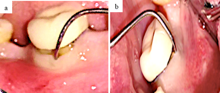
On Intra-oral clinical examination: Inflammation of marginal gingiva was seen on the buccal and lingual aspect of the teeth. A porcelain crown was seen placed on root canal treated teeth. Deep periodontal pocket was measured using Willam’s periodontal probe in relation to mesiobuccal and distolingual aspect of 36 with grade 3-furcation involvement, assessed using Naber’s probe. ([Figure 1] a,b).
Investigations
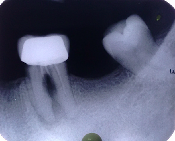
IOPA of 46 ([Figure 2]) revealed radiopacity in the occlusal surface, extending into the pulp chamber of mesial and distal roots where the mesial root was incompletely restored and perforation was seen involving the furcation area and distal root was 8mm short from the apex, suggestive of incomplete endodontic treatment. Radiolucency involving the apical area of the distal and mesial root along with radiolucency in furcation area was present.
Diagnosis
Primary endodontic with secondary periodontal lesion w.r.t. 46.
Treatment plan
• After proper evaluation and diagnosis of the condition, treatment plan was discussed with endodontist and decision was made. Informed consent was taken from patient, after explaining the need for both periodontic and re-endodontic treatment for the same tooth and understanding the importance of both treatment requirement and follow up in future.
Treatment plan follows:
Phase I: Scaling and root planing; and giving oral hygiene instructions.
Evaluation after Phase 1: Recall the patient after 1 week.
Phase II: Surgical procedure.
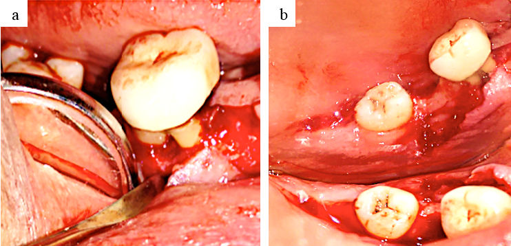
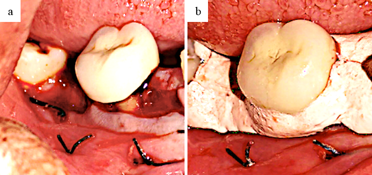
2% lignocaine hydrochloride containing adrenaline was given as local anesthesia. A full thickness mucoperiosteal flap was reflected on buccal and lingual aspect of 46 to visualize the furcation area. After thorough scaling and root debridement, with the help of tweezer and gracy curette, soft diseased tissues around the furcation area were removed. With at low-speed pear-shaped carbide bur osteoplasty and ostectomy were performed along with copious saline irrigation. A smooth and positive architecture without any bony spicules or sharp bony margins or ledges, was achieved while performing osteoplasty and ostectomy ([Figure 3]). The thickness around the flap edges was trimmed and positioned apically, using 3–0 black silk sutures, pack was placed ([Figure 4] a,b).
Oral hygiene instructions and use of 0.12% chlorhexidine digluconate mouthwash was advised to the patient. Patient was recalled after 2 weeks for suture removal.
Discussion
Bacterial contamination that has been spread from pulp to periodontium requires immediate repair and care, as it can cause serious damage to periodontium and adjacent surrounding tissues. [4] The diagnosis in this reported case was of primary endodontic with secondary periodontal involvement due to endodontic perforations caused at the time of root canal procedure, spread of bacteria from pulp to the periodontium. Endodontic treatment was administered followed by periodontal surgery after 3weeks.
A minimally invasive and conservative approach is the tunneling technique for grade III or IV furcation defects, which provide good accessibility and effective plaque control by the patient.[8]
A clear radiographic image is required to assess the bony architecture, level and extent of bone loss, root length and divergence, crown/root ratio that would help in treatment decision making. For preparation of a furcation tunnel, these factors need to be taken into consideration. The tunnel preparation is considered more effective, and success compared to other resective and regenerative surgery procedure. [10], [11], [12], [13]
Another challenge is the diagnosis and prognosis of the tooth having endo-perio lesions, which is more complex and complicating situation in today’s clinical practice. A good communication between periodontist, endodontist and patient is required for periodontal and endodontal care and patients' compliance. This would help in better understanding and highlights the importance of both procedures requirement and to ensure a correct treatment plan. [14], [15]
Conclusion
A clinician requires great skill to identify and treat a complex pathogenesis caused by Endo-Perio lesion. In this case report, a better cooperation between periodontist and endodontist is required to effectively treat the lesion, with a proper treatment plan. A few treatment options are available for treating molar teeth with severe furcation involvement, which presents a difficult challenge to the clinician. Tunnel preparation of furcation-involved molars is performed for maintaining oral hygiene, retention and functional of the affected tooth, which is considered as an effective and appropriate method for treatment of furcation involvement.
Acknowledgements
None.
Conflicts of Interest
The authors report no conflicts of interest.
Source of Funding
None.
References
- M Simring, M Goldberg. The Pulpal Pocket Approach: Retrograde Periodontitis. J Periodontol 1964. [Google Scholar] [Crossref]
- I Rotstein, R Salehrabi, J L Forrest. Endodontic treatment outcome: survey of oral health care professionals. J Endod 2006. [Google Scholar]
- L K Bakland, F M Andreasen, J O Andreasen, RE Walton, T Torabinejad. Management of traumatized teeth. Principles and Practice of Endodontics. 3rd Edn. 2002. [Google Scholar]
- N Shah. Endodontic-Periododontic Continuum. Dent Clin N A 1974. [Google Scholar]
- F M Carranza, MG Newman. Clinical Periodontology. 8th Edn. 1996. [Google Scholar]
- K P Sistla, K V Raghava, S J Narayan, U Yadalam, A Bose, P P Roy. Endo-perio continuum: A review from cause to cure�. J Adv Clin Res Insights 2018. [Google Scholar] [Crossref]
- . American Academy of Periodontology. Glossary of periodontal terms. 4th Edn.. 2001. [Google Scholar]
- A Chopra, K Sivaraman. Management of Furcal Perforation with Advanced Furcation Defect by a Minimally Invasive Tunnel Technique. Contemp Clin Dent 2018. [Google Scholar]
- A Bonaccorso, T R Tripi. Endo-perio lesion: Diagnosis, prognosis and decision-making. Endod Pract Today 2014. [Google Scholar]
- A Balachandran, S Sundaram. Resective procedures in the management of mandibular molar furcation involvement: A report of three cases. J Interdiscip Dent 2014. [Google Scholar]
- B Langer. Root resections revisited. Int J Periodontics Restor Dent 1996. [Google Scholar]
- L B Hellden, A Elliot, B Steffensen, J E Steffensen. The prognosis of tunnel preparations in treatment of class III furcations. A follow-up study. J Periodontol 1989. [Google Scholar]
- K L Lee, E F Corbet, W K Leung. Survival of molar teeth after resective periodontal therapy--a retrospective study. J Clin Periodontol 2012. [Google Scholar]
- S E Hamp, S Nyman, J Lindhe. Periodontal treatment of multirooted teeth. Results after 5 years. J Clin Periodontol 1975. [Google Scholar]
- S Nyman, B Rosling, J Lindhe. Effect of professional tooth cleaning on healing after periodontal surgery. J Clin Periodontol 1975. [Google Scholar]
