- Visibility 205 Views
- Downloads 10 Downloads
- DOI 10.18231/j.jdp.2021.030
-
CrossMark

Angina bullosahemorrhagica, an uncommon oral disorder: Case report
Introduction
Angina bullosa hemorrhagica (ABH) is an uncommon and a benign sub epithelial disorder appears as haematic blister on oral and oropharyngeal mucosa, although its real prevalence is still unknown. This condition was first described in 1933 as traumatic oral hemophlyctenosis later by Badham, in 1967 coined the term Angina bullosa hemorrhagica for this lesion. The lesions of ABH may be indistinguishable from blood blisters related to thrombocytopenia; however, blood tests and the absence of areas of ecchymosis, epistaxis, or gingival bleeding are helpful signs to rule it out. Irrespective of gender this lesion distinctively affecting adults, with the highest incidence over the 5th decade of life.[1]
The present case report describes a case of patient diagnosed with ABH and discusses the characteristics and clinical evolution of lesions, possible complications, and adequate treatment approaches, considering masticatory trauma as main etiologic agent.
Description of case
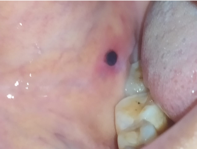
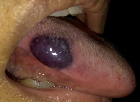
A 42-year old female patient reported at the Department of Periodontology and Oral Implantologyy, Hazaribag College of Dental Sciences and Hospital, Hazaribag, Jharkhand, India is described below. With the chief complain of sudden appearence of a blood blister on her lateral aspect of tongue while having dinner. On examination, a blood filled blister was seen on the lateral border of tongue, which was initially painless, raised round and dark red – violet in colour.
Medical and dental history was taken of the patient. Patient is under medication for type II Diabetes Mellitus (DM) since 7 years and did not give history of taking any drug or systemic agent that could affect blood coagulation (acetylsalicylic acid or blood-thinners) or any other underlying condition that could cause further systemic abnormality.
Patient was then subjected for routine blood examination, which yielded normal result.
Complete information related to the nature of the disease was explained to the patient. Non surgical treatment plan was initiated and patient was kept under observation.
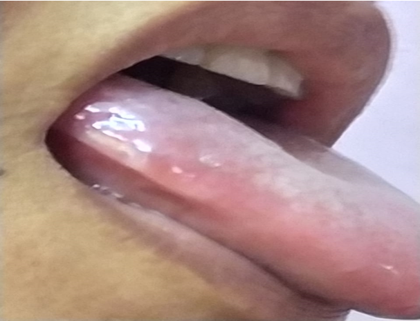
After 3 days the blister spontaneously got ruptured by itself. The patient experienced minor pain, followed by ulceration of the area, for which palliative treatment approach was done and the lesion got healed within 7 days.
|
S. No. |
Description of patient |
Data |
|
1 |
Sex |
Female |
|
2 |
Age (years) |
42 |
|
3 |
Smoking habit |
No |
|
4 |
Medical history |
Patient is under medication for diabetes since 7 years. |
|
5 |
Site affected |
Side of tongue, Buccal musoca |
|
6 |
Cause |
Dietary habit |
|
7 |
Drug treatment |
None |
|
8 |
Laboratory tests |
Bleeding time : 2.35 Clotting time : 5.32 |
|
9 |
Secondary infection |
None |
|
10 |
Treatment |
Non-surgical approach |
Discussion
ABH is characterized by the sudden appearance of a blister with blood on the oral mucosa, with no identifiable cause or related systemic disorder. While its etiology is still uncertain, it has been described as a multifactorial phenomenon, in which functional or dental trauma seem to be the most important risk factor.[2]
Traditionally, ABH is an idiopathic condition. ABH usually affect the soft palate, but these lesions can also occur in the anterior pillar of the fauces, epiglottis, arytenoids, pharyngeal wall, and esophagus.Both middle-aged and elderly individuals are more affected by ABH which ruptures spontaneously and heals without scarring. Grinspan et al. postulated the contribution of an alteration in glucose metabolism in their patients with ABH because they found DM, a positive family history of DM, or a sign of abnormal glucose metabolism in 44% of their patients. There are reports of association of ABH with hypertension, and chronic renal failure although pathogenesis is not yet cleared.[3]
In clinical practice the isolated nature of the swelling, typical clinical presentation, and rapid healing process of the blister are often sufficient to diagnose ABH. During the workup, laboratory testing, including differential blood count, and coagulation assessment tests are indicated to exclude any possible underlying hemostatic defects. A tissue biopsy or analysis of blister fluid is not advised, because it may cause a secondary infection at the blister site.[4]
Reports have suggested a possible association between ABH and systemic disorders such as diabetes[5] and hypertension,[6] although further studies aimed at confirming such preliminary findings must be performed to confirm it.
|
Ref. |
n |
Diabetes |
Hypertension |
|
Grinspan et al [8] |
54 |
24(44)% |
00 (0%) |
|
Giuliani et al [9] |
08 |
01 (12.5%) |
00 (0%) |
|
Yamamoto et al [10] |
11 |
04 (36.4%) |
03 (27.3%) |
|
Horie et al [11] |
01 |
01 (6.25%) |
06 (37.5%) |
|
Deblauwe and Van der Waal [12] |
09 |
01 (11.1%) |
00 (0%) |
|
Serra et al [13] |
04 |
00 (0%) |
02 (50%) |
|
Martini et a1 [14] |
04 |
00 (0%) |
02 (50%) |
|
Rosa et al [15] |
47 |
4 (8.5%) |
17 (32.2%) |
`
|
Disease |
Type of lesion |
Content of the blister |
Location |
Cutaneous involvement |
Involvement of other mucosal membranes |
|
Angina Bullosa hemorrhagica |
Subepithelial blister |
Hematic |
Lining Mucosa (soft palate) |
No |
Oropharynx and esophageal |
|
Mucous membrane pemphigoid |
Subepithelial blisters and vesicles |
Serous and serohematic |
Masticatory mucosa and Lining mucosa (gingiva) |
Yes |
Ocular, genital, oropharynx, nasal and esophageal |
|
Pemphigus vulgaris |
Intraepithelial blisters and vesicles |
Serous |
Masticatory mucosa and Lining mucosa (areas of friction) |
Yes |
Nasal, ocular, esophageal, genital, pharyngeal |
|
Linear IgA disease |
Subepithelial blisters and vesicles |
Serous and serohematic |
Masticatory mucosa and Lining mucosa |
Yes |
Ocular, nasal, genital |
|
Bullous amyloidosis |
Subepithelial blister |
Serous, serohematic or hematic |
Masticatory mucosa and Lining mucosa |
Yes |
Ocular, anal, vaginal, esophageal (depending on the subtype) |
Even though the diagnosis of ABH is purely clinical, the patient’s systemic condition must be considered. Singh et al point out that a history of continuous trauma of the teeth to the mucosa may lead to a presumptive diagnosis of ABH;[2] however, the differential diagnosis excludes cutaneous,
mucous or blood pathologies, such as erythema multiforme, lichen planus, pemphigus, pemphigoid, dermatitis herpetiformis, epidermolysis bullosa, oral amyloidosis and thrombocytopenia and Willebrand disease. As it is observed that blood blister in ABH spontaneously gets ruptured and heals by itself, and so no treatment is required. However benzydamine hydrochloride can be prescribed to provide symptomatic relief. [16], [17]
The first step in ABH management is patient counseling. The overall benign character of the condition and its favorable prognosis should be highlighted.
Clinical criteria for the diagnosis ofABH [18]
(For a positive diagnosis of ABH using these criteria, the case should meet a minimum of 6 out of 9 defined criteria, with criteria I and II as required)
|
I |
Clinically notable hemorrhagic bulla or erosion with a history of bleeding of the oral mucosa |
|
II |
Exclusively oral or oropharyngeal localization |
|
III |
Palate localization |
|
IV |
Triggering event or food promoting factor (food intake) |
|
V |
Recurrent lesions |
|
VI |
Favourable evolution without a scar within few days |
|
VII |
Painless lesion, tingling or burning sensation |
|
VIII |
Normal platelet count and coagulation profile |
|
IX |
Negative direct immunofluorescence |
The above mentioned table includes 9 criteria to diagnose ABH out of which 7 criteria were observed in our patient , are as follows:-
Clinically notable hemorrhagic bulla or erosion with a history of bleeding of the oral mucosa.
Exclusively oral or oropharyngeal localization.
Triggering event or food promoting factor (food intake).
Recurrent lesions.
Favourable evolution without a scar within few days.
Painless lesion, tingling or burning sensation.
Normal platelet count and coagulation profile.
Conclusion
The knowledge related to ABH in dental practice is very much necessary in order to avoid misdiagnosis, since this condition itself gets rupture and heal within 10 days of duration. It is important for the dentist to acknowledge this condition as to differentiate it from other oral vesicular processes with a poorer prognosis such as Pemphigus Vulgaris, Mucous Membrane Pemphigoid or certain hematological diseases.
Conflict of Interest
The authors declare that there are no conflicts of interest in this paper.
Source of Funding
None.
References
- S Rai, M Kaur, S Goel. Angina bullosa hemorrhagica: Report of two cases. Indian J Dermatol 2012. [Google Scholar]
- PEM Valencia, SL Otálora, MAV Valencia. Angina bullosa hemorrhagica: a case report. Rev Facultad de Odontología Universidad de Antioquia 2017. [Google Scholar] [Crossref]
- S S Arsude, B B Supekar, A S Jire. Angina bullosa hemorrhagica: A rare case report in known asthmatic on inhaled corticosteroids. Indian J Allergy 2019. [Google Scholar]
- J P Peters, P M Van Kempen, S M Robijn, H G Thomeer. Angina Bullosa Hemorrhagica: Post-traumatic Swelling in the Oral Cavity-A Case Report. J Adv Oral Res 2020. [Google Scholar]
- M Petruzzi. Angina bullosahaemorrhagica: a case report. J Biol Regul Homeost Agents 2009. [Google Scholar]
- N Horie, R Kawano, J Inaba, T Numa, T Kato, D Nasu. Angina bullosahemorrhagica of the soft palate: a clinical study of 16 cases. J Oral Sci 2008. [Google Scholar]
- A Rosa, F Geraldo Pappen, A P Neutzing Gomes. Angina bullosahemorrhagica: a rare condition?. RSBO 2012. [Google Scholar]
- N Horie, R Kawano, J Inaba, T Numa, T Kato, D Nasu. Angina bullosa hemorrhagica of the soft palate: a clinical study of 16 cases. J Oral Sci 2008. [Google Scholar]
- D Serra, H S De Oliveira, J P Reis, R Vieira, A Figueiredo, . Angina bullosa haemorrhagica: a disorder to keep in mind. Eur J Dermatol 2010. [Google Scholar] [Crossref]
- K Yamamoto, M Fujimoto, M Inoue, M Maeda, N Yamakawa, T Kirita. Angina bullosa hemorrhagica of the soft palate: report of 11 cases and literature review. J Oral Maxillofac Surg 2006. [Google Scholar] [Crossref]
- B M Deblauwe, I Van Der Waal. Blood blisters of the oral mucosa (angina bullosa haemorrhagica). J Am Acad Dermatol 1994. [Google Scholar] [Crossref]
- J Alberdi-Navarro. Angina Bullosa hemorrhagica an enigmatic oral disease. World J Stomatol 2015. [Google Scholar]
- M Z Martini, C A Lemos, E H Shinohara. Angina bullosa hemorrhagica: report of 4 cases. Minerva Stomatol 2010. [Google Scholar]
- M Giuliani, G F Favia, C Lajolo, C M Miani. Angina bullosa haemorrhagica: presentation of eight new cases and a review of the literature. Oral Dis 2002. [Google Scholar] [Crossref]
- D Grinspan, J Abulafia, H Lanfranchi. Angina bullosa hemorrhagica. Int J Dermatol 1999. [Google Scholar] [Crossref]
- B M Deblauwe, I Van Der Waal. Blood blisters of the oral mucosa (angina bullosahaemorrhagica). J Am Acad Dermatol 1994. [Google Scholar] [Crossref]
- S Edwards, J D Wilkinson, F Wojnarowska. Angina bullosahaemorrhagica--a report of three cases and review of theliterature. Clin Exp Dermatol 1990. [Google Scholar] [Crossref]
- U Ordioni, M Hadjsaïd, G Thiery, F Campana, J H Catherine, Lanr. Angina bullosahaemorrhagica: a systematic review and proposalfor diagnostic criteria. Int J Oral Maxillofac Surg 2019. [Google Scholar]