Introduction
The use of composite resins has become important in restorative dentistry.1 Currently, esthetic restorations are at the forefront of dentistry. Tooth colored restorations have become popular nowadays because of the development of materials that have better esthetic and functional features. 2 Composites comprise of properties like strength, stiffness, wear & corrosion resistance, fatigue life (long life due to repeated load) and thermal conductivity. 3
An important property of composite resin is its colour stability. In order for the esthetic restorative materials to be functional, they need to maintain colour and shade in order to blend with the neighbouring tooth structure.3 Staining or discoloration of the restorative material is one of the reasons for replacement of composite restorations which occurs because of the aging process in the oral environment induced by several extrinsic or intrinsic factors. Extrinsic factors can differ according to the individual’s nutrition, smoking and tobacco chewing habits, due to excessive intake of acidic beverages etc. Intrinsic factors include discoloration of the restorative material and depends on the resin matrix, filler weight, particle size distribution, and type of photoinititiator.1 In oral conditions, composite resins are exposed to different dietary beverages such as coffee which might result in absorption and adsorption of colorants in coffee into the resin surface and consequently undesirable color change. 4, 5
Composites selected in this study were Filtek Universal Z250XT (3M ESPE), Giomers (Beautifil Injectable and Beautifil 2). Filtek universal is a nanohybrid composite which comprises of filler content of zirconia and silica with the filler loading of 82% by weight (68% by volume). 6 It combines physical, mechanical, and esthetic properties and incorporates a high-volume fraction of filler particles with a wide particle size distribution (5-100 nm). 7, 8
Giomer is a relatively new innovative filler technology of resin composite. In place of applying purely glass or quartz as the typical fillers, the giomer encompasses inorganic fillers (ranges between 0.01 and 5 mm). 7 It is a fluoride-releasing, resin-based dental adhesive material that comprises Pre reacted glass (PRG) fillers. 9 Beautifil Injectable (Shofu) and Beautifil 2 (Shofu), both are novel composites based on Giomer technology. In a study by Tanthanuch et al. 2014, 7 Giomers showed superior color stability as compared to nanohybrid composites.
Owing to these enhanced properties, we have selected to compare and evaluate Filtek and Giomers in this study. There have not been much studies carried out and so we have chosen the materials mentioned above. Therefore, the aim of this study was to measure color stability of 3 different composite resins like Filtek Universal Z250XT (3M ESPE), Beautifil Injectable (Shofu) and Beautifil 2 (Shofu). The null hypothesis was that there the material type would not affect the color stability of resin composites.
Material and Method
Composite specimen preparation
For preparation of specimens, three different light‑cured resin composites were used and grouped as follows, with 10 samples in each group (n=10):
Group 1: Filtek 3M ESPE,
30 samples (n=10) were prepared by placing them in the 2-mm deep and 5-mm internal diameter plastic rings, interplaced between two glass slides and pressed, allowing for a smooth surface and no gap formation. The specimens were then light‑cured for 40 seconds using the exit window of a quartz‑tungsten‑halogen light polymerization unit that was placed against the glass slab. The specimens were stored under moist conditions at 37oC until the radiographic part of the experiment was conducted.
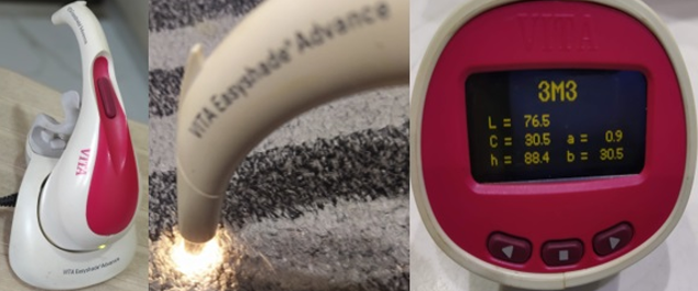
Specimens of all 3 groups (total 30) were immersed in distilled water at 37°C for 24 hours. Following the first immersion cycle, all specimens were removed, rinsed under tap water and blot-dried for initial colorimetric measurement with a spectrophotometer (Vita Easyshade, Germany). [Figure 1]
Colorimetric measurements of the specimens were performed according to the CIELab color scale, recording the L*, a*, and b* values, where L* is the lightness coordinate, and a* and b* are the chromacity coordinates in the red-green axis and the yellow-blue axis, respectively (2.1). Three readings were performed and a mean value for the L*, a*, and b* values was obtained for each specimen. Later, all the specimens were kept immersed in staining solution (Green Tea, Tetley) for 1 week which was prepared by mixing 25 g of powder in 250 ml of water which was standardized and changed every day.
Color measurements were recorded after 1 week of immersion. One examiner performed all the recordings of the L*, a*, and b* values. ΔE’ values were calculated using Hunter’s equation, ΔE’=[(ΔL*)2+(Δa*)2+(Δb*)2]1/2.
To determine the relationship between the amount of color alteration recorded on a spectrophotometer to the clinical environment, data were converted to the National Bureau of Standards (NBS) system. According to this system, ΔE values can be described by the subsequent equation: NBS unit = ΔE × 0.92. [Table 1]
Results
According to the values obtained from Table 1, National Bureau of Standards (NBS) values of the composites were evaluated based on their color alteration.
Filtek: 11.75 (Remarkably apparent alteration), Beautifil Injectable: 9.23 (Remarkably apparent alteration), Beautifil 2: 5.97 (Apparent alteration).
There were statistically significant differences among the restorative materials when the results were compared using the one-way ANOVA (P < 0.05) [Table 2].
There was statistically significant difference found in Filtek after staining on 7th day (P value: 0.004) indicating its least color stability, followed by Beautifil Injectable which was not statistically significant (P value: 0.101). Beautifil 2 showed least color change, thereby proving color stable on the 7th day, however it was not statistically significant (P value: 0.609) [Table 3].
According to NBS system, at the end of 7th day, Filtek (11.75) showed remarkably apparent alteration, followed by Beautifil Injectable which also showed remarkably apparent alteration (9.23) and Beautfil 2 which showed only Apparent alteration (5.97).
Table 1
National Bureau of Standards (NBS) system of expressing color differences
Table 2
One way analysis of variance results
|
Score |
Sum of squares |
df |
Mean Square |
F |
Sig (p value) |
|
Between groups |
412.06 |
5 |
82.41 |
9.795 |
0.000 |
|
Within groups |
201.93 |
24 |
8.41 |
|
|
|
Total |
613.999 |
29 |
|
|
|
Discussion
Color stability of dental restorations is one of the most important characteristics of composite resin materials in terms of longevity.4 Resin composites can be subjected to a variety of sources of staining during their lifespan. Among the frequent sources is the dietary consumption of staining beverages. So, the inherent property of the material, such as color stability is very important in order to maintain an esthetic result.3 Thus, the objective of the current investigation was to compare three esthetic resin composite formulations in regard to their color stability. Green Tea solution (Tetley) has been used in the current study as it is frequently consumed throughout the world. 3
Shade changes before and after immersion in staining solution was measured using a spectrophotometer in order to yield the CIEL*a*b* parameters. 3 The established CIE Lab system, one of the most common color measurement systems in dentistry today, was used in this study with precise results for several color parameters.10, 11 Here, L* or lightness variable is proportional to “value” in the Munsell system and a* and b* variables are chromacity coordinates designating red/green and yellow/blue axes respectively. This method is able to detect subtle changes in shades of dental resins and it is a widely used approach to objectively determine color changes in dental restorations expressed as numerical values without the inherent subjectivity of the operator decision-making process. 12
The ΔE value presents relative color differences of dental materials or tooth surfaces before and after an intervention. According to literature, values of ΔE < 1 are regarded as not appreciable by the human eye. Values 1 < ΔE < 3.3 are considered appreciable by skilled operators but clinically acceptable, whereas values of ΔE > 3.3 are considered appreciable by nonskilled persons and are, hence, not clinically acceptable. Therefore, color changes above a value of ΔE = 3.3 were considered clinically unacceptable. 4
Materials tested were Filtek, Beautifil Injectable and Beautifil 2. Filtek comprises of filler content of zirconia and silica with the filler loading of 82% by weight (68% by volume). The later 2 comprise of Giomer technology. Giomer is a fluoride-releasing, resin-based dental adhesive material that comprises PRG fillers. PRG fillers are fabricated by the acid-base reaction between fluoroalumino –silicate glass (FASG) and polyalkenoic acid (PAA) in the presence of water to form a wet siliceous hydrogel. After freeze-drying, the desiccated xerogel was further milled and silanized to form PRG fillers of a specific size range. 9, 13
The benefits of S-PRG filler are: release ions other than fluoride, that serve a range of benefits, (i) Fluoride release and fluoride recharge, (ii) formation of acid resistant layer (iii) reinforcement of tooth structure (iv) antiplaque effect (v) remineralization of dentin (vi) acid buffering capacity and reduce acid production by acidogenic bacteria. 9, 14
Owing to these enhanced properties, we have selected Giomers in our study. Beautifil 2 showed the highest color stability after 1 week followed by Beautifil Injectable and the least stability was observed in Filtek. Moreover, Poggio et al. 15 in their study have reported that Filtek formulation was susceptible to color changes.3 This can be attributed to the fact that the resin matrix of the giomer restoratives were all based on Bis-GMA/TEGDMA mixtures. Bis-GMA molecules are highly viscous and require the addition of low molecular weight monomers to achieve a workable consistency upon filler incorporation. TEGDMA is often employed as the diluent monomer for Bis-GMA due to its low viscosity and excellent copolymerization characteristics. The lower staining susceptibility of the Beautifil 2 and Beautifil Injectable giomers may be attributed in part to the relatively higher TEGDMA content when compared to their conventional counterparts. 12, 16 Additionally, in a study by Hajira et al. (2015) 9 the enhanced property of color stability of giomers was elaborated in addition to various other properties like fluoride release and recharge, ion release & modulation effect, buffering of lactic acid, antibacterial properties, adherence of streptococcus mutans, PRG and biofilm and mineral induction. Hence, as there were significant differences between the materials tested, the null hypothesis of this study was rejected.
Conclusion
Among the materials tested, Filtek Z250 showed the highest color alteration and Beautifil II showed the lowest highest color alteration. In clinical practice when selecting an appropriate restorative material, the dentists should consider the drinking and oral hygiene habits of the patients and the factors that may affect the color stability of restorative materials. In addition, patients should also be informed about the staining potential of the restorations.


