- Visibility 468 Views
- Downloads 26 Downloads
- Permissions
- DOI 10.18231/j.jdp.2021.040
-
CrossMark
- Citation
Alignment of rotated permanent maxillary central incisors with segmental orthodontics in mixed dentition: A case report
- Author Details:
-
Jaya Verma
-
Vipin Ahuja *
Abstract
Segmental Orthodontics is an essential twig of Pediatric Orthodontics being practiced nowadays in growing children in mixed dentition worldwide. Early interception of malocclusion either involves full arch or a segment of arch to be treated for malocclusion; and when a segment is used, it is calculated as ‘Segmental Orthodontics’. It’s a simplified approach in interceptive orthodontics as it involves only a segment of few teeth which is more children friendly. The present case report displays derotation of maxillary central incisors in a male child with simplified segmental orthodontics in mixed dentition period. The results obtained were satisfactory and rapid. Therefore, this technique can be widely applied to treat such type of mixed dentition malocclusions.
Introduction
Interceptive pediatric orthodontics envelopes the protocol required to treat either developed mal-alignments or to prevent the severity of developed malocclusions. Early interception either involves full arch or a segment of arch to be treated for malocclusion; and when a segment is used, it is considered as ‘Segmental Orthodontics’. [1], [2] Segmental Orthodontics in children is advantageous in many ways over full arch orthodontics. It’s a simplified approach in pediatric orthodontics as it involves only a segment of few teeth. It is also psychologically acceptable to the growing children and is less time consuming. It is successfully used to treat various incipient and moderate malocclusions of mixed and permanent dentition. The cost incurred is significantly less and is an established management approach in managing varied malocclusions.[3]
There is enough literature documented before to shore up the fact that early interception of malocclusion in mixed dentition is of paramount importance and should be dealt with precedence. Most common early-age malocclusions observed nowadays in practice are single tooth cross-bites and anterior teeth displacements due to mesiodens, other supernumerary teeth or odontomes, derotations etc. The treatment protocol used can vary from ‘2x6 appliance’, ‘2x4 appliance’ or ‘Brackets on few teeth with NITI approach’. [3]
Tooth rotation is considered subjectively as any evident (at least 20°) mesiolingual or distobuccal intra-alveolar displacement of tooth around its longitudinal axis. [4] The prevalence of tooth rotation is found to be 2.1-5.1% in the untreated population,. [5] The rotation of permanent teeth can be divided into two groups based on etiologic factors: [6], [7], [8]
Rotation of permanent teeth due to pre-eruptive disturbances: Injury of the pre-maxillary region in childhood, that displaced and misaligned the developing tooth bud, the presence of an adjacent pathology such as cyst, tumor, odontoma, supernumerary tooth (mesiodens) can cause rotations of teeth.
Rotation of permanent teeth due to post-eruptive disturbances: There are habitual, mechanical, local, or environmental factors such as space availability for tooth alignment, path of tooth eruption and functional effects produced by tongue and lips, can cause rotations of teeth.
The treatment of maxillary anterior permanent teeth with rotation can be performed by several methods: The routine treatment of rotated teeth is use of fixed appliance either full arch fixed therapy or segmental arch therapy. Also, in some conditions such as slight rotation, correction can be performed with a removable appliance such as an appliance with a labial bow and palatal spring. The Pediatric dentist is a specialized dentist who come across these young malocclusions at a very young age and should have a sound clinical knowledge to diagnose and correct these malalignments with the simple protocols like segmental orthodontics in pediatric dentistry.
The present case shows the treatment of rotated central incisors with ‘Brackets on few teeth with NITI approach’
Case Report
A 8 year old boy reported to the Department of Pediatric and Preventive Dentistry, Hazaribagh College of Dental Sciences and Hospital with a chief complaint of severe rotations in upper front two teeth since 2 years ([Figure 1]).
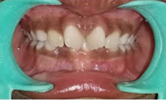
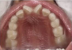
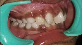
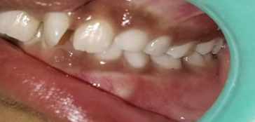
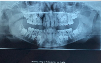
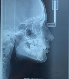
History did not reveal any previous trauma in that region. Extra oral examination in a lateral view displayed patient with slightly convex profile and competent lips and in frontal view, he was mesoprosopic. Intraoral examination revealed vital 11 and 21 with mesiopalatal rotaions ([Figure 2]). Mandibular occlusion was noted as Angle Class I with crowding in lower anterior teeth region with more than 70° rotation of right and left maxillary central incisors ([Figure 3], [Figure 4]). Orthopentamogram revealed both the central incisors were young permanent teeth with a ¾ of root completion in relation to 21 and root almost completed in relation to 11. In the radiographic examination of OPG and lateral cephalogram ([Figure 5], [Figure 6]), pathologic problems such as supernumerary teeth or odontoma were not discovered; skeletal relationship of the patient was Class I with mandibular plane angle in a nominal range. Alginate impressions of both arches were taken to fabricate study and analyze the study models for space, and no space deficiency was found. Rotated teeth showed early stage of root development and there was enough space in the upper arch to derotate it.
Treatment
Segmental orthodontics was planned from first primary molar of right side to first primary molar of left involving a segment of 8 teeth without using primary second molars and permanent molars. Roth 0.018 prescription brackets were used and placed on 11,12,53,54,21,22,63 and 64 as per standardized prescription protocol. 12 NiTi wire was engaged in the brackets with elastic modules ([Figure 7], [Figure 8]).
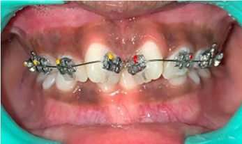
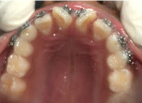
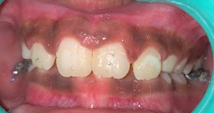
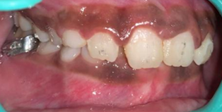
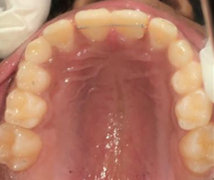
The lingual arch space maintainer was planned and given for the mandibular arch crowding of lower anteriors and maintenance of arch length. The patient was called after one month of bracket and wire placement and significant derotation was observed with the maxillary central incisors; the 12 NITI wire was continued till gross rotation was corrected with 11 and 21. The wire sequence followed after this was 14 NITI, 16 NITI and 16SS and the brackets were debonded after 6 months ([Figure 9], [Figure 10]). The fixed permanent lingual retainer with 11 and 21 was bonded at the cingulum level ([Figure 11]). The child and parents were happy with the results and patient was recalled after 3 months for follow-up.
Discussion
Tooth rotation, is defined as observable mesiolingual or distolingual intra-alveolar displacement of the tooth around its longitudinal axis. [5], [7] Gupta et al.[4] classified the rotation into three groups: <45°, 45-90° and >90°. The findings of this study inferred that rotations were the most common (10.24%) type of anomalies among the study population and the majority of tooth rotations were between 45° and 90°, followed by <45° rotations. Mandibular second premolars are most commonly rotated followed by mandibular first premolars and maxillary central incisors with the same occurrence
There are two types of orthodontic treatments for intercepting teeth rotations: removable and fixed. The removable treatment approach for correction of tooth rotation involves removable appliance with a labial bow or palatal springs like z-spring to derotate the teeth. The major limitation with removable appliances is that it can only correct mild rotations of less than 45° and also indicated in the case of maxillary central incisors rotaions. This method is unable to derotate other teeth, severe rotations and multiple teeth rotations. In addendum to this, rotations are also associated with a very high risk of relapse and as patient compliance is a factor in the removable appliance, relapse even in the treatment phase is more likely to happen.[9]
The fixed treatment approach for correction of tooth rotation is the use of fixed arch or segmental orthodontic therapy. This protocol is advantageous as it does need patient compliance and is a controlled and efficient method for derotations. However, the limitations are time required and oral hygiene maintenance with full arch therapies. There is paucity in literature to highlight the use of segmental orthodontics in the management of tooth rotations. Few studies document 2 x 4 and 2x6 appliances as a successful treatment approach for derotations of teeth in the mixed dentition.[6] Although this approach can correct all kind of rotations, but use of it has some limitations. One of the major limitations cited in the text is that these appliances can be used only after complete eruption of permanent first molars and incisors while in some situations where immediate correction of rotated tooth become necessary, they are not indicated.[10] So, we have used a simplified approach of using 8 teeth for fixed orthodontic therapy without involving first permanent molars. And the result outcome was satisfactory.
Treatment of derotations at a very early stage of mixed dention is recommended by many authors. Mavragani et al. advised to initiate orthodontic correction of the incisors at a young age during mixed dentition as an introductory phase of treatment; reason given was root shortening of incisors due to apical resorption associated with orthodontic treatment.[11] Before any derotation treatment is undertaken, sufficient space is to be checked for the availablity to accommodate the tooth in alignment.[12] Relapse is not uncommon in derotations. The high risk of relapse is due to stretching of the supra-alveolar and transseptal gingival fibers, which readapt very slowly to the new position. Therefore, it should be overcorrected or long term retention is given after finishing the treatment. [6]
Conclusions
This segmental orthodontic approach can be effectively used in the mixed dentition for treating rotations of permanent incisors relatively in a short duration.
This protocol is simplified approach (without involving permanent molars) to treat minor orthodontic corrections in mixed dentition.
Segmental orthodontics is an outstanding option to treat rotated teeth.
Conflict of Interest
The authors have stated explicitly that there are no conflicts of interest in connection with this article.
Source of Funding
None
References
- Agarwal A, Mathur. Segmental Orthodontics for the Correction of Cross Bites Int. J Clin Ped Dent. 2011;4(1):43-7. [Google Scholar]
- Verma J, Ahuja V. Interception of developing anterior malocclusion due to supernumerary tooth by “2 x 4 Appliance”: A clinical case report. J Dent Panacea. 2021;3(1):40-5. [Google Scholar]
- Ahuja V. Segmental Orthodontics: Simplified approach in Pediatric Orthodontics . J Dent Panacea. 2021;3(3):97-8. [Google Scholar]
- Gupta SK, Saxena P, Jain S, Jain D. Prevalence and distribution of selected developmental dental anomalies in an Indian population. J Oral Sci. 2011;53(2):231-8. [Google Scholar] [Crossref]
- Shpack N, Geron S, Floris I, Davidovitch M, Brosh T, Vardimon AD. Bracket placement in lingual vs labial systems and direct vs indirect bonding. Angle Orthod. 2007;77(3):509-17. [Google Scholar] [Crossref]
- Kim YH, Shiere FR, Fogels HR. Pre-eruptive factors of tooth rotation and axial inclination. J Dent Res. 1961;40(3):548-57. [Google Scholar]
- Parisay I, Boskabady M, Abdollahi M, Sufiani M. Treatment of severe rotations of maxillary central incisors with whip appliance: Report of three cases. Dent Res J (Isfahan). 2014;11(1):133-9. [Google Scholar]
- Baccetti T. Tooth rotation associated with aplasia of nonadjacent teeth. Angle Orthod. 1998;68(5):471-4. [Google Scholar] [Crossref]
- Jalaly T, Jahanbin A, Ahrari F. Introduction to removable orthodontic appliances and dentofacial orthopedics. Mashhad: Vajhegane Kherad. . 2007. [Google Scholar]
- Hess E, Campbell PM, Honeyman AL, Buschang PH. Determinants of enamel decalcification during simulated orthodontic treatment. Angle Orthod. 2011;81(5):836-42. [Google Scholar] [Crossref]
- Mavragani M, Bøe OE, Wisth PJ, Selvig KA. Changes in root length during orthodontic treatment: Advantages for immature teeth. Eur J Orthod. 2002;24(1):91-7. [Google Scholar] [Crossref]
- Isaacson KG, Muir JD, Reed RT. Removable Orthodontic Appliances. 2nd Edn.. . 2003. [Google Scholar]
How to Cite This Article
Vancouver
Verma J, Ahuja V. Alignment of rotated permanent maxillary central incisors with segmental orthodontics in mixed dentition: A case report [Internet]. J Dent Panacea. 2021 [cited 2025 Oct 08];3(4):185-189. Available from: https://doi.org/10.18231/j.jdp.2021.040
APA
Verma, J., Ahuja, V. (2021). Alignment of rotated permanent maxillary central incisors with segmental orthodontics in mixed dentition: A case report. J Dent Panacea, 3(4), 185-189. https://doi.org/10.18231/j.jdp.2021.040
MLA
Verma, Jaya, Ahuja, Vipin. "Alignment of rotated permanent maxillary central incisors with segmental orthodontics in mixed dentition: A case report." J Dent Panacea, vol. 3, no. 4, 2021, pp. 185-189. https://doi.org/10.18231/j.jdp.2021.040
Chicago
Verma, J., Ahuja, V.. "Alignment of rotated permanent maxillary central incisors with segmental orthodontics in mixed dentition: A case report." J Dent Panacea 3, no. 4 (2021): 185-189. https://doi.org/10.18231/j.jdp.2021.040
