- Visibility 20 Views
- Downloads 5 Downloads
- DOI 10.18231/j.jdp.2023.039
-
CrossMark
- Citation
Prosthetic rehabilitation of hemisected tooth using Pier Abutment: A beacon of hope for decaying tooth
Introduction
During the past decade, dentistry has experienced a paradigm shift from its past to newer concepts. Improved and highly conservative treatment modalities have been introduced, which has increased patient’s preference to the save teeth that would have earlier been planned for extraction. These improved techniques have given the patient an opportunity to preserve and maintain their functional dentition.[1] Supposedly, if the tooth is affected partially then hemisection procedure can be performed and the decayed part is removed and the sound tooth structure remains in place. Hemisection basically means sectioning of a mandibular molar into two halves subsequently removal of the diseased root and its coronal portion.[2] Here the retained part of the tooth is endodontically treated and the furcation area is made self-cleansable by removing the lip of root carefully, followed by its rehabilitation with an extracoronal restoration.[3] For an apt selection of cases, Periodontal, Prosthodontic, and Endodontic evaluation is crucial.[4] From a periodontal point of view, hemisection is recommended when there is class III furcations involvement or severe bone loss limited to one root, if the patient faces difficulty to perform proper oral hygiene in the area or an extensive exposure of the roots due to dehiscence. On the other hand from a restorative perspective, hemisection can be performed when a portion of the tooth can be retained to act as the abutment for the prosthesis or tooth having vertical root fracture limited to single root of multirooted tooth, any destructive process limited to single root of a multirooted tooth like trauma, caries or external root resorption. Contraindication for the procedure is the presence of fused roots or proximity of the roots which can lead to difficulty in its separation. [5] Present case report enlightens a simple process of hemisection in mandibular molar and its subsequent restoration and rehabilitation.
Case Report
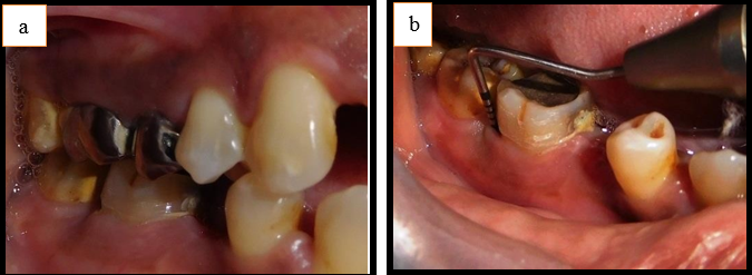
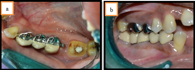
A 35-year-old female patient reported to the department of Prosthodontics and crown & bridge with a chief complaint of pain in her lower right back region of jaw since one and half months. Pain was dull and irregular in nature, which usually aggravated on mastication and the other complaint was of missing tooth in the same region of jaw. On extraoral examination, no other abnormality seen. While on intraoral examination, it was seen that lower right mandibular first molar was sensitive to percussion along with grade I mobility and there was missing 45 in the same region. On probing the area, periodontal pocket of 11 mm was found on the buccal and distal surfaces of 46 along with grade II furcation ([Figure 1]a,b).
Patient was advised to get the IOPA of the concerned region. IOPA confirmed the presence of grade II furcation defect together with loss of periodontal bone along the distal root of 46. Periodontal bone support was found to be good on the mesial root of 46. Interdental bone loss was noticed between 46 and 47 region. Patient was diagnosed with chronic generalized gingivitis accompanying localized periodontitis with respect to 46. The appropriate treatment comprises extraction with respect to 46 followed by prosthetic rehabilitation of 45, 46 with removable partial denture, fixed partial denture or implant. Since, patient was not willing for extraction, a conservative approach was chosen which consisted hemisection of distal root of 46 followed by prosthetic rehabilitation of 45,46.
Treatment Procedures
Endodontic Phase
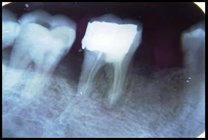
Intentional root canal treatment was done with respect to 46. After 14 days of obturation, patient was adviced to get hemisection done with respect to distal root of 46 ([Figure 2]).
Periodontic phase
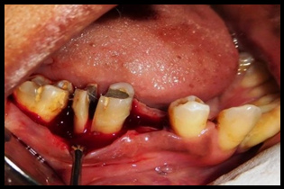
Hemisection was done with respect to distal root of 46 in the following manner. At first, Right inferior alveolar, long buccal and lingual nerve block was given using 2% lignocaine. After that, crevicular incision was made from lower right first premolar region to lower right second molar region then full thickness mucoperiosteal flap was raised to provide an access for proper visualization, instrumentation and minimal surgery. After opening of the flap, both curettage and debridement was done. Vertical cut was made facio-lingually along the bifurcation area using the long shank tapered fissure carbide bur ([Figure 3]) and the distal root was extracted ([Figure 4]).
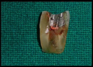
Extreme care was taken not to alter the underlying bone and its adjacent structures while removing the distal portion of the root. Using betadine solution, irrigation of the extracted socket was done. Then to remove the developmental ridges odontoplasty was done while distal aspect of the mesial root was contoured for facilitating oral hygiene measures.
Socket preservation was done by placing “Bio-Oss” in the extraction socket. 3-0 mersilk sutures were placed and COE pack dressing was given at the surgical site ([Figure 5]).
.
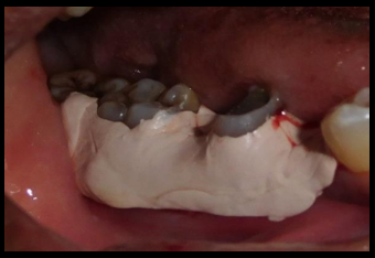
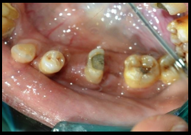
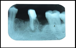
The surgical site was kept undisturbed for atleast 4 weeks with no occlusal stress on the mesial root aspect of 46, so as to have better healing of the surgical site. Patient was recalled after 3 months, surgical site was evaluated clinically, good post hemisection healing was seen ([Figure 6]) and IOPA was taken which revealed good bone translation ([Figure 7]). After that prosthetic rehabilitation of the hemisected tooth was planned with fixed partial denture (pier abutment) with respect to mesial root of 46 and 44,45,47.
Prosthodontic phase
After 3 months of complete healing of the surgical site, prosthetic rehabilitation of fixed partial denture using pier abutment with respect to mesial root of 46 and 44,45,47 was done in the following manner.
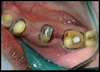
Firstly preliminary impressions were made of maxillary and mandibular arch using irreversible hydrocolloid impression material and then casts were obtained. Facebow record was made using springbow and transferred to the semi-adjustable articulator (hanau wide-vue II) and the maxillary cast was mounted on it. Then using interocclusal record mandibular diagnostic cast was mounted to evaluate any type of occlusal prematurities, accordingly occlusal equilibration was done. Tooth preparation was done with respect to 44, mesial part of 46, 47 ([Figure 8]) and then gingival retraction was done.
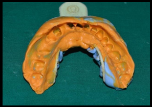
Final impression was made using addition silicone elastomeric impression material by putty-reline technique and the master cast was obtained ([Figure 9]).
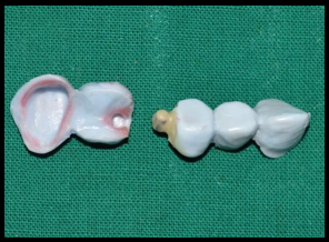
Provisional restoration was made using tooth colored selfcure acrylic resin and then placed. Using interocclusal record mandibular master cast was mounted. The wax pattern was fabricated using pattern wax for mandibular right first premolar, second premolar region and mesial half of mandibular first molar after that female counterpart of pier abutment was cut to fit the dovetail on distal aspect of pier abutment ([Figure 10]).
Then surveying was done to evaluate the parallelism of dovetail; dovetail female was then placed within the correct contour of pier abutment and then casting of the female portion was done and excess height of female portion was cut down. Male pattern was seated in the casted female portion, then wax pattern was fabricated of distal part of mandibular first molar and second molar. The mandrel was cut off from male pattern and its casting was done.
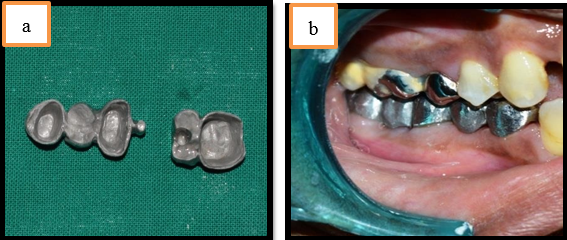
Metal coping trial was done to evaluate the proper seating of female and male counterparts of pier abutment ([Figure 11]a,b)
Then ceramic facing was added to the metal coping having anterior segment with female portion and posterior segment with male portion which were then assembled together and checked for appropriate seating of the prosthesis intraorally.
During cementation, anterior segment with keyway was cemented first followed by cementation of posterior segment having key using glass ionomer cement ([Figure 12]a,b).
Post cementation instructions were given concerning its maintenance and proper oral hygiene. To evaluate the healing and success of the restoration periodic recall was done.
Discussion
For an apt selection of cases, Periodontal, Prosthodontic, and Endodontic evaluation is crucial. [4] Buhler stated that, before performing any molar extraction it should undergo hemisection procedure as it gives better, biological and cost saving alternative along with long term sucess. [6] Variable treatment alternatives to rehabilitate damaged and unrestorable tooth includes dental implant, fixed partial denture and removable partial denture. The aim here is to preserve what is present by performing hemisection, which ultimately provides a prognosis similar to any other tooth with endodontic treatment.
Endodontic phase
At first, the endodontic treatment is performed the reason behind this is if in case endodontic treatment cannot be performed on this tooth or endodontic treatment failed then hemisection procedure will be contraindicated in such cases.
Periodontic phase
Some crucial factors to be considered for choosing molar for hemisection procedure are as follows:
Root divergence: The hemisected root should have greater amount of root divergence, since root proximity cause difficulty in performing hemisection.
Root form: Mandibular molars commonly shows concavity on its distal roots therefore it is very important to perform odontoplasty so as to achieve appropriate contour.
Location of furcation: Furcation opening should be present closer to the cement-enamel junction so as to have better prognosis of the retained roots.
Remaining root attachment: it is very difficult assess; because long, ovoid, cylindrical roots serves as an outstanding abutment. [7]
Prosthodontic phase
Immediately upon loosening the part of root support from a tooth, it needs a restoration that allows the tooth to function independently or to act as an abutment for the fixed partial denture. Accordingly, restoration is necessary for appropriate function and stabilization of occlusion. Some requirements to be taken into considerations during the prosthesis fabrication: A taper of more than 6-10 degrees should be given to the hemisected abutment so as to achieve path of insertion compatible with anterior abutment along with that it compensates the buccal and lingual grooves placed in the abutment. [8] To reduce the lateral forces and eliminate the non-working contacts the cuspal inclines are made less steeper. Stein said, “whenever considering esthetics, for the posterior region sanitary pontic can be considered as the best choice.” [9] Implants can be considered as the best choice with appropriate function but in above case as concern with the patient’s requirement and economical barrier, the procedure of hemisection of the molar and its prosthetic rehabilitation proved to be successful. [10]
Conclusion
Appropriate planning, operative sequence along with multidisciplinary approach applied in the present case emphasized the importance of specialized knowledge and professional communication. Hemisection is a boon for extracting teeth, if done with all the appropriate majors leading to the longterm success of the procedure.
Author’s Contribution
Data gathering: Dr. Sreenivasarao Ghali, Dr. Bindu S Patil
Study design and writing: Dr. Dishita Chokhani, Dr. Kiran Ghatole.
Editing and approval of final draft: Dr. Sreenivasarao Ghali, Dr. Dishita Chokhani.
Conflict of Interest
There is no conflict of interest.
Source of Funding
None.
References
- A Buragohain, A Nagpal, V Bhatia, H Kaur. Functional rehabilitation of hemisected mandibular molar: A case report. J Dent 2019. [Google Scholar]
- WJ Kost, JE Stakiw. Root amputation and hemisection. J Can Dent Assoc 1991. [Google Scholar]
- RH Rapoport, P Deep. Traumatic hemisection and restoration of a maxillary first premolar: a case report. Gen Dent 2003. [Google Scholar]
- U Radke, R Kubde, A Paldiwal. Hemisection: A window of hope for freezing tooth. Case Rep Dent 2012. [Google Scholar] [Crossref]
- FS Weine. . Endodontic Therapy. 5th edn 1996. [Google Scholar]
- SF Rosenstiel, MF Land, J Fujimoto. . Contemporary fixed prosthodontics. 2nd edn. 1995. [Google Scholar]
- G Parmar, P Vashi. Hemisection: a case report and review. Endodontology 2003. [Google Scholar]
- M Desanctis, KG Murphy. The role of resective periodontal surgery in the treatment of furcation defects. Periodontol 2000. [Google Scholar] [Crossref]
- MS Schmitt, HF Brown. The hemisected mandibular molar: A strategic abutment. J Prosthetic Dent 1987. [Google Scholar]
- MN Saad, J Moreno, C Crawford. Hemisection as an alternative treatment for decayed multirooted terminal abutment: A Case Report. J Can Dent Assoc 2009. [Google Scholar]
