Introduction
The term dentigerous refers to ‘tooth bearing’. Dentigerous cyst being the most common developmental cyst of odontogenic origin contributes to 49% of all cystic lesions. It develops around the coronal portion of an unerupted tooth.1
It is formed due to the obstruction of venous flow by the developing tooth and fluid collection between the follicular epithelium and the crown of the developing or unerupted tooth.2 It has a higher male predilection. 1, 3
The three types of dentigerous cysts: central, lateral, or circumferential are based on their relationship to the involved tooth crown. The central form appears around the crown, the lateral variant partially covers the crown and the circumferential form encompasses both the coronal and the radicular portion of the involved tooth. 4
The most common site is the mandibular third molar (77%), followed by the maxillary third molar (11%), and canine (5%), and only 1% were related to impacted supernumerary teeth. The highest incidence is noted in the second to fourth decades. 2
In this case report, we present a unique, unusual, and extensive case of a dentigerous cyst involving the mandible region crossing the midline and associated with a supernumerary tooth.
Case History
A 48-year-old male patient reported with a chief complaint of swelling in the right lower back tooth region since 2 years. The patient gave history of pain and loss of sensation since two months.
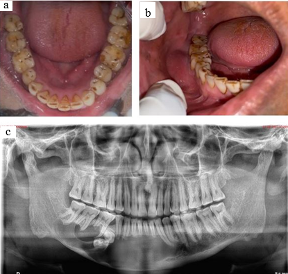
Intraoral examination revealed missing 45 [Figure 1a] and a well-defined dome-shaped swelling in the buccal vestibule in relation to 44 to 48 region measuring approximately 2x2 cm. [Figure 1b] On palpation, expansion of the buccal cortex was noted from 43 to 48 region with vestibular obliteration and tenderness noted from 34 to 48 region.
A provisional diagnosis of odontogenic cyst and differential diagnoses of dentigerous cyst and ameloblastoma were considered.
Investigations
Fine Needle aspiration cytology was suggestive of an acute inflammatory lesion.
The orthopantomogram revealed a well-defined radiolucent lesion extending from the distal aspect of 34 to 48 region with smooth hydraulic borders with root resorption of the involved tooth and impacted 45 and a partially developed supernumerary tooth inside the lesion. [Figure 1c]
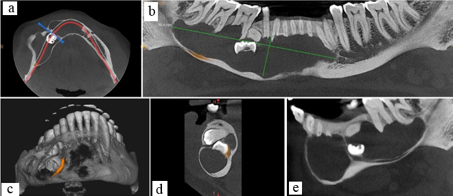
Cone Beam Computed Tomography revealed a well-defined radiolucent lytic lesion extending from the distal aspect of 34 to the distal aspect of 47 regions measuring approximately 85.4x21.3x18.8mm (A-P, S-I, M-D). [Figure 1a] The internal structure showed septations with impacted 45 and supernumerary tooth. Scalloped borders and knife-edged pattern of root resorption noted in relation to the involved teeth.
Expansion and perforation of buccal cortex with thinning of lingual cortex was noted. The inferior alveolar nerve was displaced along the intact thinned-out lower border of mandible. [Figure 1b,c&2d]
Cystic enucleation and extraction of 31 to 47 along with endodontic treatment of 32, 33, and 34 was done.
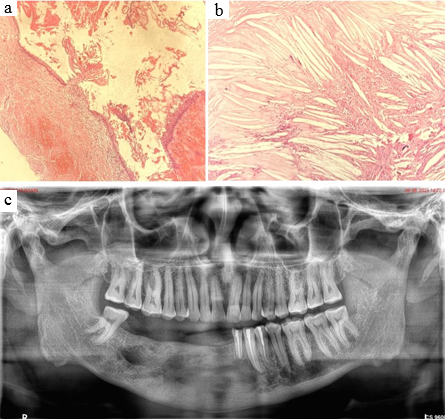
The histopathological section showed multiple tissue bits cyst lined by stratified squamous epithelium with focal acanthosis and absence of rete ridges, suggestive of a dentigerous cyst.[Figure 1a & b]
One-year follow-up and radiographs were taken and the patient was asymptomatic [Figure 1c]
Figure 1
a: Intraoral picture showing missing 45; b: Intraoral picture of the swelling; c: Orthopantomogram of the lesion
Click here to view

Figure 2
a: Axial section (CBCT); b: Panoromic reconstruction showing the extend of the lesion; c: 3D reconstruction section; d & e: Saggital section (CBCT)
Click here to view

Figure 3
a: Histopathological image showing cyst wall lined by stratified squamous epithelium; b: Wall of the cyst displaying cholesterol clefts with foreign body giant cell reaction; c: Orthopantomogram after one –year follow up
Click here to view

Discussion
The present case highlights a dentigerous cyst arising from a partially developed supernumerary tooth. Dentigerous cysts which form from the reduced enamel epithelium encapsulate coronal portion of teeth. 2, 4
Proliferation of cyst causes expansion leading to the release of bone-resorbing factors resulting in increased osmolality of the cystic fluid. These changes can cause facial asymmetry, tooth displacement, and compression of vital structures. In our case, the inferior alveolar nerve bundle was compressed and the patient presented with paraesthesia.5, 6
Multiple dentigerous cysts are related to Marotreux-Lamy syndrome and cleidocranial dysplasia.3, 7
Clinically dentigerous cyst is asymptomatic and is identified as an accidental radiography finding.8
Radiographically dentigerous cyst presents as a symmetrical unilocular radiolucency around the crown of a tooth.2, 4
In our case the lesion was completely radiolucent with septations and impacted 45 and a supernumerary tooth and the lesion seemed to arise from the crown of the supernumerary tooth.
Histologically, the lumen is bordered by 2 to 4 cell layers of cuboidal to flattened non-keratinized epithelial cells that may generate keratin by metaplasia.4
The choice of treatment depends on various factors like age, size, location, root developmental stage, and relationship to the adjacent tooth. Enucleation and marsupialization are the treatment options for dentigerous cysts.9, 10 In our case complete removal of the cyst was performed.
The lining epithelium of dentigerous cysts is pluripotent and can turn into mural ameloblastoma, mucoepidermoid carcinoma, or squamous cell carcinoma.2 Therefore when the cyst is completely removed, the prognosis is better, and recurrence is uncommon.

